Trichothecium roseum
| Trichothecium roseum | |
|---|---|
 | |
| Scientific classification | |
| Kingdom: | Fungi |
| Division: | Ascomycota |
| Subdivision: | Pezizomycotina |
| Class: | Sordariomycetes |
| Order: | Hypocreales |
| Family: | Incertae sedis |
| Genus: | Trichothecium |
| Species: | T. roseum |
| Binomial name | |
| Trichothecium roseum (Pers.) Link (1809) | |
| Synonyms | |
| |
Trichothecium roseum is a fungus in the division Ascomycota first reported in 1809.[1] It is characterized by its flat and granular colonies which are initially white and develop to be light pink in color.[1] This fungus reproduces asexually through the formation of conidia with no known sexual state.[1] Trichothecium roseum is distinctive from other species of the genus Trichothecium in its characteristic zigzag patterned chained conidia.[2] It is found in various countries worldwide and can grow in a variety of habitats ranging from leaf litter to fruit crops.[2] Trichothecium roseum produces a wide variety of secondary metabolites including mycotoxins, such as roseotoxins and trichothecenes, which can infect and spoil a variety of fruit crops.[1] It can act as both a secondary and opportunistic pathogen by causing pink rot on various fruits and vegetables and thus has an economical impact on the farming industry.[1] Secondary metabolites of T. roseum, specifically Trichothecinol A, are being investigated as potential anti-metastatic drugs. Several agents including harpin, silicon oxide, and sodium silicate are potential inhibitors of T. roseum growth on fruit crops.[3][4][5] Trichothecium roseum is mainly a plant pathogen and has yet to show a significant impact on human health.[1]
History and classification
The genus Trichothecium is small and heterogeneous comprising seventy-three recorded species.[1] This genus was first reported in 1809.[1] The main members of the genus include Trichothecium polybrochum, Trichothecium cystosporium, Trichothecium pravicovi, and Trichothecium roseum.[1] Trichothecium roseum has morphologically different conidiophores and conidia than the other three main species, which made development of these features the center of extensive study throughout the years.[1] Since Trichothecium fungi lack a sexual phase, systematic classification was not uniform following their discovery.[1] These fungi were initially grouped into Fungi imperfecti under the form classification Deuteromycetes.[1] In 1958, Tubaki expanded Hughes’ classification of soil Hyphomycetes, part of the form class of Fungi imperfecti, by adding a ninth section in order to accommodate T. roseum and its unique conidial apparatus.[6][7] Trichothecium has now been classified under the class Sordariomycetes.[1] A recent classification has placed Trichothecium under the phylum Ascomycota since they produce conidial stages that are similar to the perfect fungi.[1]

Morphology
Trichothecium roseum colonies are flat, granular, and powdery in appearance.[1][2] The color of the colonies appears to be white initially and develop into a light pink to peach color.[1] The genus Trichothecium is characterized by its pinkish colored colonies.[8]
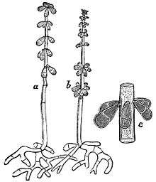
Conidiophores of T. roseum are usually erect and are 200-300μm in length.[9] They arise singly or in loose groups.[1] Conidiophores are simple hyphae,[10] which are septate in their lower half,[6] and bear clusters of conidia at the tip.[2] These conidiophores are indistinguishable from vegetative hyphae until production of the first conidium.[1] Conidium development is distinctive[2] and was first described by Ingold in 1956.[6] Conidia arise as blowouts from the side of the conidiophore apex which is thus incorporated into the base of each spore.[6] After the first conidium is blown out, before it matures, the apex of the conidiophore directly below blows out a second conidium from the opposite side.[6] Conidia are pinched out from the conidiophore one after another in alternating directions in order to form the characteristic zigzag patterned chain.[1] Conidia of T.roseum (15-20 × 7.5-10 μm)[9] are smooth and clavate.[1] Each conidium is two celled with the apical cell being larger than the curved basal cell.[1] Conidia are light pink and appear translucent under the microscope.[1] They appear a more saturated pink colour when grown in masses in culture or on the host surface.[1]
Growth and physiology
Trichothecium roseum reproduces asexually by the formation of conidia with no known sexual stage.[1] Trichothecium roseum is relatively fast-growing as it can form colonies reaching 9 cm (4 in) in diameter in ten days at 20 °C (68 °F) on malt extract agar.[8] This fungus grows optimally at 25 °C (77 °F) with a minimum and maximum growing temperature of 15 °C (59 °F) and 35 °C (95 °F) respectively.[8] Trichothecium roseum can tolerate a wide pH range but grows optimally at a pH of 6.0. Sporulation occurs rapidly at pH 4.0-6.5 and a combination of low temperature (15 °C (59 °F)) and high glucose concentration can increase the size of conidia.[8] Treatment of T. roseum with colchicine increases the number of nuclei in conidia, growth rate, and biosynthetic activities.[8] There are a variety of sugars that T. roseum can utilize including D-fructose, sucrose, maltose, lactose, raffinose, D-galactose, D-glucose, arabinose, and D-mannitol.[8] Good growth also occurs in the presence of various amino acids including L-methionine, L-isoleucine, L-tryptophan, L-alanine, L-norvaline, and L-norleucine.[8]
Secondary metabolites
Trichothecium roseum can produce numerous secondary metabolites that include toxins, antibiotics, and other biologically active compounds.[1] Diterpenoids produced include rosolactone, rosolactone acetate, rosenonolactone, desoxyrosenonolactone, hydroxyrosenonolactones, and acetoxy-rosenonolactone. Several sesquiterpenoids are also produced by T. roseum including crotocin, trichothecolone, trichothecin, trichodiol A, trichothecinol A/B/C, trichodiene, and roseotoxin.[1][8][11]
Biomedical applications
Trichothecium roseum was found to antagonize pathogenic fungi, such as Pyricularia oryzae (Magnaporthe oryzae) and Phytophthora infestans, in vitro.[12] It was suggested that the antifungal compound trichothecin was the main contributor to this action.[12] In other studies trichothecinol B isolated from T. roseum displayed modest antifungal activity against Cryptococcus albidus and Saccharomyces cerevisiae.[13]
Various studies have indicated that Trichothecinol A isolated from T. roseum strongly inhibited TPA-induced tumour promotion on mouse skin in carcinogenesis tests and therefore may be valuable for further investigation as cancer preventive agent.[13][14][15] Anti-cancer studies have also shown that Trichothecinol A significantly inhibits cancer cell migration and therefore can be developed as a potential new anti-metastatic drug.[15]
Habitat and ecology
Trichothecium roseum is a saprophyte [10] and is found worldwide.[8] It has been found in soils in various countries including Poland, Denmark, France, Russia, Turkey, Israel, Egypt, the Sahara, Chad, Zaïre, central Africa, Australia, Polynesia, India, China, and Panama.[8] Known habitats of T. roseum include uncultivated soils, forest nurseries, forest soils under beech trees, teak, cultivated soils with legumes, citrus plantations, heathland, dunes, salt-marshes, and garden compost.[8] Commonly, this fungus can be isolated from the tree leaf litter of various trees including birch, pine, fir, cotton, and palm.[8] It has also been isolated from several food sources such as barley, wheat, oats, maize, apples, grapes, meat products, cheese, beans, hazelnuts, pecans, pistachios, peanuts, and coffee.[10] Levels of T. roseum in foods other than fruits are generally low.[10]
Plant pathology
There are approximately two hundred twenty-two different plant hosts of T. roseum found worldwide.[1] Trichothecium roseum causes pink rot on various fruits and vegetables.[1] It is considered both a secondary and opportunistic pathogen since it tends to enter the fruit/vegetable host through lesions that were caused by a primary pathogen.[1] Disease caused by this fungus is characterized by the development of white powdery mold that eventually turns pink.[1] Antagonistic behaviours of T. roseum with certain plant pathogenic fungi was reported by Koch in 1934.[16] He started that T. roseum actively parasitized stroma of Dibotryon morbosum which causes black knot disease in cherry, plum, and apricot trees.[16]
Apple disease
Trichothecium roseum is known to produce pink rot on apples particularly following an apple scab infection caused by Venturia inaequalis.[1] Studies have shown that roseotoxin B, a secondary metabolite of T. roseum, can penetrate apple peels and cause lesions.[17] Trichothecium roseum also causes apple core rot which is a serious problem in China.[18] Core rot not only causes economic loss but it is also associated with high levels of mycotoxin production.[18] There have been reports of the presence of trichothecenes, specifically T-2 toxin, in infected apples in China.[18] T-2 toxin has the highest toxicity of the trichothecenes and poses a threat to individuals who consume these infected apples due to its carcinogenicity, neurotoxicity, and immunotoxicity.[18]
Grape disease
Trichothecium roseum was identified, along with Acremonium acutatum, as the two strains of pathogenic fungi which caused white stains on harvested grapes in Korea.[19] The presence of mycelia on the surface of the grapes resulted in a white stained, powdery mildew appearance.[19] Trichothecium roseum was identified using fungal morphology and nucleotide sequencing by PCR.[19] It appears as though the fungus covers the surface of the grape only and does not penetrate into the tissue.[19] This stain lowers the quality of the grapes and causes serious economic losses.[19]
Trichothecin, trichothecolone, and rosenonolactone, which are secondary metabolites of T. roseum, were detected in wines.[20] Presence of small quantities of trichothecin can inhibit alcohol fermentation.[20] Trichothecium roseum rot has been reported to be increasing in wineries in Portugal.[20] In this case, T. roseum appeared to grow over rotten grapes that were infected with gray rot.[20] Mycotoxins were only detected in wines that were made with grapes that had gray rot and thus these toxins may be indicators of poor quality grapes.[20] Grape contamination by T. roseum appears to be prominent in temperate climates.[20]
Other fruit disease
Cases of T. roseum pink rot have been reported on numerous other fruits, however detailed studies have yet to be pursued.[1] Pink T. roseum rot has been reported on tomatoes in Korea and Pakistan.[21][22] It also causes pink rot in muskmelons and watermelons in Japan, the United States, South America, India, and the United Kingdom.[1] Trichothecium roseum is reported to grow also on bananas and peaches.[1]
Prevention of plant disease
Preventative measures can be taken to avoid growth of T. roseum in fruit crops.[23] These include ensuring adequate ventilation in the storage facility, avoiding injuring and bruising the fruit, and ensuring adequate storage temperatures.[23] Pre- and postharvest applications have been suggested as measures to control T. roseum production on fruit crops.[1] In particular, studies have been done on testing various compounds to prevent T. roseum growth on several melon types.[3][4][5] Harpin was inoculated on harvested Hami melons and caused significantly reduced lesion diameter and thus decreased T. roseum growth.[3] Silicon oxide and sodium silicate also reduced the severity of pink rot and lesion diameter in harvested Hami melons.[4] Pre-harvest inoculation of harpin on muskmelons decreased the amount of pink rot caused by T. roseum on harvested melons.[5]
References
- 1 2 3 4 5 6 7 8 9 10 11 12 13 14 15 16 17 18 19 20 21 22 23 24 25 26 27 28 29 30 31 32 33 34 35 Batt, C.A.; Tortorello, M (2014). Encyclopedia of food microbiology (2 ed.). London: Elsevier Ltd. p. 1014. ISBN 978-0-12-384730-0.
- 1 2 3 4 5 Onions, A.H.S.; Allsopp, D.; Eggins, H.O.W. (1981). Smith's introduction to industrial mycology (7th ed.). London, UK: Arnold. ISBN 0-7131-2811-9.
- 1 2 3 Yang, B; Shiping, T; Jie, Z; Yonghong, G (2005). "Harpin induces local and systemic resistance against Trichothecium roseum in harvested Hami melons". Postharvest Biology and Technology. 38 (2): 183–187. doi:10.1016/j.postharvbio.2005.05.012.
- 1 2 3 Guo, Y; Liu, L; Zhao, J; Bi, Y (2007). "Use of silicon oxide and sodium silicate for controlling Trichothecium roseum postharvest rot in Chinese cantaloupe (Cucumis melo L.)". International Journal of Food Science & Technology. 42 (8): 1012–1018. doi:10.1111/j.1365-2621.2006.01464.x.
- 1 2 3 Wang, J; Bi, Y; Wang, Y; Deng, J; Zhang, H; Zhang, Z (2013). "Multiple preharvest treatments with harpin reduce postharvest disease and maintain quality in muskmelon fruit (cv. Huanghemi)". Phytoparasitica. 42 (2): 155–163. doi:10.1007/s12600-013-0351-8.
- 1 2 3 4 5 Barron, George L. (1968). The genera of Hyphomycetes from soil. Baltimore, MD: Williams & Wilkins. ISBN 9780882750040.
- ↑ Kendrick, W. B.; Cole, G. T. (1969). "Conidium ontogeny in hyphomycetes and its meristem arthrospores". Canadian Journal of Botany. 47 (2): 345–350. doi:10.1139/b69-047.
- 1 2 3 4 5 6 7 8 9 10 11 12 Domsch, K.H.; Gams, Walter; Andersen, Traute-Heidi (1980). Compendium of soil fungi (2nd ed.). London, UK: Academic Press. ISBN 9780122204029.
- 1 2 Watanabe, Tsuneo (2009). Pictorial atlas of soil and seed fungi : morphologies of cultured fungi and key to species (3rd ed.). Boca Raton, Fla.: CRC. ISBN 978-1-4398-0419-3.
- 1 2 3 4 Pitt, J.I.; Hocking, A.D. (1999). Fungi and food spoilage (2nd ed.). Gaithersburg, Md.: Aspen Publications. ISBN 0834213060.
- ↑ Cole, Richard; Jarvis, Bruce; Schweikert, Milbra (2003). Handbook of secondary fungal metabolites. Oxford: Academic. ISBN 978-0-12-179460-6.
- 1 2 Zhang, XiaoMei; Li, GuoHong; Ma, Juan; Zeng, Ying; Ma, WeiGuang; Zhao, PeiJi (9 January 2011). "Endophytic fungus Trichothecium roseum LZ93 antagonizing pathogenic fungi in vitro and its secondary metabolites". The Journal of Microbiology. 48 (6): 784–790. doi:10.1007/s12275-010-0173-z.
- 1 2 Konishi, Kazuhide; Iida, Akira; Kaneko, Masafumi; Tomioka, Kiyoshi; Tokuda, Harukuni; Nishino, Hoyoku; Kumeda, Yuko (June 2003). "Cancer preventive potential of trichothecenes from Trichothecium roseum". Bioorganic & Medicinal Chemistry. 11 (12): 2511–2518. doi:10.1016/S0968-0896(03)00215-3.
- ↑ Iida, Akira; Konishi, Kazuhide; Kubo, Hiroki; Tomioka, Kiyoshi; Tokuda, Harukuni; Nishino, Hoyoku (December 1996). "Trichothecinols A, B and C, potent anti-tumor promoting sesquiterpenoids from the fungus Trichothecium roseum". Tetrahedron Letters. 37 (51): 9219–9220. doi:10.1016/S0040-4039(96)02187-9.
- 1 2 Taware, R; Abnave, P; Patil, D; Rajamohananan, P; Raja, R; Soundararajan, G; Kundu, G; Ahmad, A (2014). "Isolation, purification and characterization of Trichothecinol-A produced by endophytic fungus Trichothecium sp. and its antifungal, anticancer and antimetastatic activities". Sustainable Chemical Processes. 2 (1): 8. doi:10.1186/2043-7129-2-8.
- 1 2 Freeman, G.G.; Morrison, R.I. (1949). "Metabolic products of Trichothecium roseum Link". Biochemical Journal. 45 (2): 191–199.
- ↑ Žabka, Martin; Drastichová, Kamila; Jegorov, Alexandr; Soukupová, Julie; Nedbal, Ladislav (July 2006). "Direct Evidence of Plant-pathogenic Activity of Fungal Metabolites of Trichothecium roseum on Apple". Mycopathologia. 162 (1): 65–68. doi:10.1007/s11046-006-0030-0.
- 1 2 3 4 Tang, Y; Xue, H; Bi, Y; Li, Y; Wang, Y; Zhao, Y; Shen, K (2014). "A method of analysis for T-2 toxin and neosolaniol by UPLC-MS/MS in apple fruit inoculated with Trichothecium roseum". Food Additives & Contaminants: Part A. 32 (4): 480–487. doi:10.1080/19440049.2014.968884.
- 1 2 3 4 5 Oh, S.Y.; Nam, K.W.; Yoon, D.H. (2014). "Identification of and isolated from Grape with White Stain Symptom in Korea". Mycobiology. 42 (3): 269–273. doi:10.5941/MYCO.2014.42.3.269.
- 1 2 3 4 5 6 Serra, R; Braga, A; Venâncio, A (2005). "Mycotoxin-producing and other fungi isolated from grapes for wine production, with particular emphasis on ochratoxin A". Research in Microbiology. 156 (4): 515–521. doi:10.1016/j.resmic.2004.12.005.
- ↑ Han, K.S.; Lee, S.C.; Lee, J.S.; Soh, J.W. (2012). "First Report of Pink Mold Rot on Tomato Fruit Caused by Trichothecium roseum in Korea". Research in Plant Disease. 18 (4): 396–398. doi:10.5423/RPD.2012.18.4.396.
- ↑ Hamid, M.I.; Hussain, M; Ghazanfar, M.U.; Raza, M; Liu, X.Z. (2014). "Causes Fruit Rot of Tomato, Orange, and Apple in Pakistan". Plant Disease. 98 (9): 1271–1271. doi:10.1094/PDIS-01-14-0051-PDN.
- 1 2 Rees, D; Farrell, G; Orchard, J.E. (2006). Crop post-harvest : science and technology. Oxford: Blackwell Science. p. 464. ISBN 978-0-632-05725-2.
External links
| Wikispecies has information related to: Trichothecium roseum |
| Wikimedia Commons has media related to Trichothecium roseum. |