Central retinal vein
| Central retinal vein | |
|---|---|
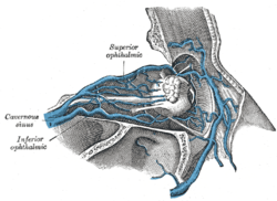
 Veins of orbit. (Central retinal vein not labeled, but region is visible - the vein is inside the optic nerve.) | |
 Diagram of the blood vessels of the eye, as seen in a horizontal section. (Central retinal vein not labeled, but region is visible. The central retinal vein is at bottom running away from the retina through the optic nerve.) | |
| Details | |
| Drains to |
Superior ophthalmic vein, cavernous sinus |
| Artery | Central retinal artery |
| Identifiers | |
| Latin | Vena centralis retinae |
| MeSH | Central+retinal+vein |
| Dorlands /Elsevier | v_05/12849780 |
| TA | A12.3.06.111 |
| FMA | 51799 |
The central retinal vein (retinal vein) is a short vein that runs through the optic nerve, leaves the optic nerve 10 mm from the eyeball and drains blood from the capillaries of the retina into either superior ophthalmic vein or into the cavernous sinus directly. The anatomy of the veins of the orbit of the eye varies between individuals, and in some the central retinal vein drains into the superior ophthalmic vein, and in some it drains directly into the cavernous sinus.[1][2]
Pathology
The central retinal vein is the venous equivalent of the central retinal artery, and like that blood vessel can suffer from occlusion (central retinal vein occlusion), similar to that seen in ocular ischemic syndrome.
References
- ↑ Venous Anatomy of the Orbit Cheung and McNab. Investigative Ophthalmology and Visual Science. 2003;44:988-995
- ↑ MeSH entry for central retinal vein - National Library of Medicine - Medical Subject Headings - 2007
Additional images
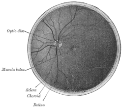
 Interior of posterior half of bulb of left eye. The veins are darker in appearance than the arteries.
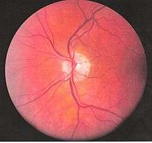
 Eye vessels
This article is issued from
Wikipedia.
The text is licensed under Creative Commons - Attribution - Sharealike.
Additional terms may apply for the media files.