Periapical cyst
| Periapical cyst | |
|---|---|
 | |
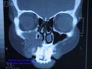
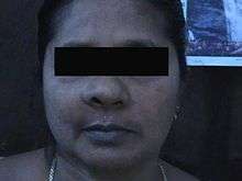
| CT scan through head showing a right periapical cyst | |
| Classification and external resources | |
| Specialty | endodontics |
| ICD-10 | K09.0 |
| DiseasesDB | 31994 |
| eMedicine | ent/681 |
The periapical cyst (also termed radicular cyst or dental cyst) is the most common odontogenic cyst. It is caused by pulpal necrosis secondary to dental caries or trauma. The cyst lining is derived from the cell rests of Malassez. Usually, the periapical cyst is asymptomatic, but a secondary infection can cause pain. On radiographs, it appears a radiolucency (dark area) around the apex of a tooth's root.
Signs and symptoms

Expansion of the cyst causes erosion of the floor of the maxillary sinus. As soon as it enters the maxillary antrum the expansion starts to occur a little faster because there is space available for expansion. Tapping the affected teeth will cause shooting pain. This is virtually diagnostic of pulpal infection.
Radiographically it is virtually impossible to differentiate granuloma from a cyst. If the lesion is large it is more likely to be a cyst. Radiographically both granuloma and cyst appear radiolucent, associated with the apex of non vital tooth.
Causes
Dental cysts are usually caused due to root infection involving the tooth affected greatly by carious decay . The resulting pulpal necrosis causes release of toxins at the apex of the tooth leading to periapical inflammation. This inflammation leads to the formation of reactive inflammatory (scar) tissue called periapical granuloma further necrosis and damage stimulates the Malassez epithelial rests, which are found in the periodontal ligament, resulting in the formation of a cyst that may be infected or sterile (The epithelium undergoes necrosis and the granuloma becomes a cyst). These lesions can grow into large lesions because they apply pressure over the bone causing resorption . The toxins released by the breakdown of granulation tissue is one of the common causes of bone resorption.
These cysts are not true neoplasms
Treatment
The source (i.e., necrotic pulp) should be removed by full pulpectomy (i.e., root canal therapy) or extraction of the offended tooth, and the cyst should be enuclated.
See also
References
- Kahn, Michael A. Basic Oral and Maxillofacial Pathology. Volume 1. 2001.
- DelBalso AM (1995). "Lesions of the jaws". Semin. Ultrasound CT MR. 16 (6): 487–512. PMID 8747414. doi:10.1016/S0887-2171(06)80022-3.
- Harris M (1974). "A review of recent experimental work on the dental cyst" (Scanned image & PDF). Proc. R. Soc. Med. 67 (12 Pt 1): 1259–63. PMC 1645893
 . PMID 4449871.