Naegleriasis
| Naegleriasis | |
|---|---|
| Synonyms | primary amoebic meningoencephalitis (PAM), amebic encephalitis, naegleria infection, amoebic meningitis |
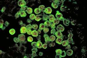 | |
| Histopathology of primary amebic meningoencephalitis due to Naegleria fowleri. Direct fluorescent antibody stain. | |
| Pronunciation |
|
| Classification and external resources | |
| Specialty | Infectious disease |
| ICD-10 | A06.6, B60.2 |
| ICD-9-CM | 136.2 |
Naegleriasis (also known as primary amoebic meningoencephalitis) is an infection of the brain by the free-living unicellular Naegleria fowleri.
N. fowleri is typically found in warm bodies of fresh water, such as ponds, lakes, rivers, and hot springs. It is also found in soil, poorly maintained municipal water supplies, water heaters, near warm-water discharges of industrial plants, and in poorly chlorinated or unchlorinated swimming pools, in an amoeboid or temporary flagellate stage. There is no evidence of it living in salt water.
Although infection occurs rarely,[1] it nearly always results in death,[2] with a case fatality rate greater than 95%.[3]
Signs and symptoms
Onset of symptoms one to nine days following exposure (with an average of five).[4] Initial symptoms include changes in taste and smell, headache, fever, nausea, vomiting, and a stiff neck. Secondary symptoms include confusion, hallucinations, lack of attention, ataxia, and seizures. After the start of symptoms, the disease progresses rapidly over three to seven days, with death occurring usually from seven to fourteen days later,[5] although it can take longer. In 2013, a man in Taiwan died twenty-five days after being infected by Naegleria fowleri.[6]
It affects healthy children or young adults who have recently been exposed to bodies of fresh water.[7] Some people have presented with a clinical triad of edematous brain lesions, immune suppression, and fever.[8]
Cause
N. fowleri invades the central nervous system via the nose, specifically through the olfactory mucosa of the nasal tissues. This usually occurs as the result of the introduction of water that has been contaminated with N. fowleri into the nose during activities such as swimming, bathing, or nasal irrigation.
The amoeba follows the olfactory nerve fibers through the cribriform plate of the ethmoid bone into the skull. There, it migrates to the olfactory bulbs and subsequently other regions of the brain, where it feeds on the nerve tissue, resulting in significant necrosis and bleeding.[9]
The organism then begins to consume cells of the brain, piecemeal, by means of an amoebostome, a unique actin-rich sucking apparatus extended from its cell surface.[10] It then becomes pathogenic, causing primary amoebic meningoencephalitis (PAM or PAME).
Pathogenesis
Naegleria fowleri propagates in warm, stagnant bodies of freshwater (typically during the summer months), and enters the central nervous system after insufflation of infected water by attaching itself to the olfactory nerve.[7] It then migrates through the cribriform plate and into the olfactory bulbs of the forebrain,[11] where it multiplies itself greatly by feeding on nerve tissue.
Diagnosis
N. fowleri can be grown in several kinds of liquid axenic media or on non-nutrient agar plates coated with bacteria. Escherichia coli can be used to overlay the non-nutrient agar plate and a drop of cerebrospinal fluid sediment is added to it. Plates are then incubated at 37 °C and checked daily for clearing of the agar in thin tracks, which indicate the trophozoites have fed on the bacteria.[12] Detection in water is performed by centrifuging a water sample with E. coli added, then applying the pellet to a non-nutrient agar plate. After several days, the plate is microscopically inspected and Naegleria cysts are identified by their morphology. Final confirmation of the species' identity can be performed by various molecular or biochemical methods.[13] Confirmation of Naegleria presence can be done by a so-called flagellation test, where the organism is exposed to a hypotonic environment (distilled water). Naegleria, in contrast to other amoebae, differentiates within two hours into the flagellate state. Pathogenicity can be further confirmed by exposure to high temperature (42 °C): Naegleria fowleri is able to grow at this temperature, but the nonpathogenic Naegleria gruberi is not.
Prevention
Michael Beach, a recreational waterborne illness specialist for the Centers for Disease Control and Prevention, stated in remarks to the Associated Press that wearing of nose-clips to prevent insufflation of contaminated water would be effective protection against contracting PAM, noting that "You'd have to have water going way up in your nose to begin with".[14]
Treatment
Since its first description in the 1960s, only seven people worldwide have been reported to have survived PAM as of 2015, with three in the United States and one in Mexico.[15][16] The prognosis remains poor for those who contract PAM, and survival remains less than 1%.[7]
On the basis of the laboratory evidence and case reports, amphotericin B has been the traditional mainstay of PAM treatment since the first reported survivor in the United States in 1982.[16][17]
Treatment has often also used combination therapy with multiple other antimicrobials in addition to amphotericin, such as fluconazole, miconazole, rifampicin and azithromycin. They have shown limited success only when administered early in the course of an infection.[18] Fluconazole is commonly used as it has been shown to have synergistic effects against naegleria when used with amphotericin in-vitro.[16]
While the use of rifampicin has been common, including in all four North American cases of survival, its continued use has been questioned.[16] It only has variable activity in-vitro and it has strong effects on the therapeutic levels of other antimicrobials used by inducing cytochrome p450 pathways.[16]
Steroids such as dexamethasone have also been used to try to reduce inflammation of the brain.[19]
In 2013, the two most recent successfully treated cases in the United States utilized drug combinations that included the medication miltefosine as well as targeted temperature management.[15] There is currently no data on how well miltefosine is able to reach the central nervous system.[16] The U.S. CDC is currently offering miltefosine to doctors for the treatment of free-living ameobas including naegleria.[15]
Untimely diagnoses remain a very significant impediment to the successful treatment of infection, as most cases have only been discovered post mortem. Infection killed 121 people in the United States from 1937 through 2007.
Epidemiology
The disease is rare and highly lethal: there have only been 300 cases as of 2008.[20] Drug treatment research at Aga Khan University in Pakistan has shown that in-vitro drug susceptibility tests with some FDA approved drugs used for non-infectious diseases have proved to kill Naegleria fowleri with an amoebicidal rate greater than 95%.[21] The same source has also proposed a device for drug delivery via the transcranial route to the brain.[22]
This form of nervous system infection by amoeba was first documented in Australia in 1965.[23][24] In 1966, four cases were reported in the USA. By 1968 the causative organism, previously thought to be a species of Acanthamoeba or Hartmannella, was identified as Naegleria. This same year, occurrence of sixteen cases over a period of two years (1963–1965) was reported in Ústí nad Labem, Czechoslovakia.[25] In 1970, the species of amoeba was named N. fowleri.[26]
The number of cases of infection could increase due to climate change.[27] In 2016, an infection was contracted in Maryland, four miles south of the Pennsylvania border;[28] this was the northernmost [29] North American case other than the three or four Minnesota cases from 2008 to 2015.[30] Additionally, the numbers of reported cases are expected to show an increase, simply because of better informed diagnoses being made both in living patients and also in autopsy findings.[31]
History
Australian physicians Fowler and Carter first described human disease caused by amebo-flagellates in Adelaide in 1965.[32][33] Their work on amebo-flagellates has provided an example of how a protozoan can effectively live both freely in the environment, and in a human host. Since 1965, more than 144 cases have been confirmed in different countries. In 1966, Fowler termed the infection resulting from N. fowleri, primary amoebic meningoencephalitis (PAM) to distinguish this central nervous system (CNS) invasion from other secondary invasions made by other amoebae such as Entamoeba histolytica.[32] A retrospective study determined the first documented case of PAM possibly occurred in Britain in 1909.[34]
The specific name, Naegleria fowleri, was named for Mathieu Naegler (1867–1934), a French parasitologist and zoologist who discovered the species for the first time in 1899 and Malcolm Fowler (1924–1974), the Australian doctor who described the distinct disease process of the pathogen in Australia in 1965.
Society and culture
Naegleria fowleri is also known as the "brain-eating amoeba." The term "brain-eating amoeba" has also been applied to Balamuthia mandrillaris, causing some confusion between the two; Balamuthia mandrillaris is unrelated to Naegleria fowleri, however, and causes a different disease called granulomatous amoebic encephalitis. Unlike naegleriasis, which is usually seen in people with normal immune function, granulomatous amoebic encephalitis is usually seen in people with poor immune function, such as those with HIV/AIDS or leukemia.[35]
Research
The U.S. National Institutes of Health budgeted $800,000 for research on the disease in 2016.[36]
Phenothiazines have been tested in vitro and in animal models of PAM.[37]
See also
References
- ↑ "The Centers for Disease Control and Prevention, Division of Parasitic Diseases – Naegleria fowleri - Primary Amoebic Meningoencephalitis (PAM) - General Information". Retrieved 2014-05-26.
- ↑ "6 die from brain-eating amoeba after swimming". MSNBC. Associated Press. 28 September 2007.
- ↑ Cetin N, Blackall D (Apr 2012). "Naegleria fowleri meningoencephalitis". Blood. 119 (16): 3658. PMID 22645743. doi:10.1182/blood-2011-06-353136.
- ↑ "Illness & Symptoms | Naegleria fowleri | CDC". www.cdc.gov.
- ↑ "CDC - 01 This Page Has Moved: CDC Parasites Naegleria". Retrieved 27 July 2015.
- ↑ Su MY, Lee MS, et al. (Apr 2013). "A fatal case of Naegleria fowleri meningoencephalitis in Taiwan". Korean J Parasitol. 51 (2): 203–6. PMC 3662064
 . PMID 23710088. doi:10.3347/kjp.2013.51.2.203.
 - 1 2 3 Centers for Disease Control and Prevention (CDC) (2008). "Primary amebic meningoencephalitis – Arizona, Florida, and Texas, 2007". MMWR. Morbidity and mortality weekly report. 57 (21): 573–7. PMID 18509301.
- ↑ Mayer, Peter (2011). "Amebic encephalitis". Surgical Neurology International. 50 (2): 50. doi:10.4103/2152-7806.80115.
- ↑ Gautam PL, Sharma S; et al. (Jan 2012). "A rare case of survival from primary amebic meningoencephalitis". Indian Crit Care med. 16 (1): 34–6. PMC 3338237 . PMID 22557831. doi:10.4103/0972-5229.94432.
- ↑ Marciano-Cabral, F; John, DT (1983). "Cytopathogenicity of Naegleria fowleri for rat neuroblastoma cell cultures: scanning electron microscopy study". Infection and immunity. 40 (3): 1214–7. PMC 348179 . PMID 6852919.
- ↑ Cervantes-Sandoval I, Serrano-Luna Jde J, García-Latorre E, Tsutsumi V, Shibayama M (September 2008). "Characterization of brain inflammation during primary amoebic meningoencephalitis". Parasitol. Int. 57 (3): 307–13. PMID 18374627. doi:10.1016/j.parint.2008.01.006.
- ↑ Donald C. Lehman; Mahon, Connie; Manuselis, George (2006). Textbook of Diagnostic Microbiology (3rd ed.). Philadelphia: Saunders. ISBN 1-4160-2581-2.
- ↑ Pougnard, C.; Catala, P.; Drocourt, J.-L.; Legastelois, S.; Pernin, P.; Pringuez, E.; Lebaron, P. (2002). "Rapid Detection and Enumeration of Naegleria fowleri in Surface Waters by Solid-Phase Cytometry". Applied and Environmental Microbiology. 68 (6): 3102–7. PMC 123984 . PMID 12039772. doi:10.1128/AEM.68.6.3102-3107.2002.
- ↑ "6 die from brain-eating amoeba in lakes", Chris Kahn/Associated Press, 9/28/07
- 1 2 3 "Naegleria fowleri — Primary Amebic Meningoencephalitis (PAM) — Amebic Encephalitis". April 23, 2015. Retrieved 17 January 2016.
- 1 2 3 4 5 6 Grace, Eddie; Asbill, Scott; Virga, Kris (2015-11-01). "Naegleria fowleri: Pathogenesis, Diagnosis, and Treatment Options". Antimicrobial Agents and Chemotherapy. 59 (11): 6677–6681. ISSN 0066-4804. PMC 4604384 . PMID 26259797. doi:10.1128/AAC.01293-15.
- ↑ Seidel, James S.; Harmatz, Paul; Visvesvara, G. S.; Cohen, Arthur; Edwards, Jack; Turner, Jerrold (1982-02-11). "Successful Treatment of Primary Amebic Meningoencephalitis". New England Journal of Medicine. 306 (6): 346–348. ISSN 0028-4793. PMID 7054710. doi:10.1056/NEJM198202113060607.
- ↑ Bauman, Robert W. (2009). "Microbial Diseases of the Nervous System and Eyes". Microbiology, With Diseases by Body System (2nd ed.). San Francisco: Pearson Education. p. 617.
- ↑ Linam, W. Matthew; Ahmed, Mubbasheer; Cope, Jennifer R.; Chu, Craig; Visvesvara, Govinda S.; Silva, Alexandre J. da; Qvarnstrom, Yvonne; Green, Jerril (2015-03-01). "Successful Treatment of an Adolescent With Naegleria fowleri Primary Amebic Meningoencephalitis". Pediatrics. 135 (3): e744–e748. ISSN 0031-4005. PMC 4634363 . PMID 25667249. doi:10.1542/peds.2014-2292.
- ↑ Caruzo G, Cardozo J (October 2008). "Primary amoebic meningoencephalitis: a new case from Venezuela". Trop Doct. 38 (4): 256–7. PMID 18820207. doi:10.1258/td.2008.070426.
- ↑ Mannan Baig Abdul; Kulsoom Huma; Ahmed Khan Naveed (2014). "Primary amoebic meningoencephalitis: amoebicidal effects of clinically approved drugs against Naegleria fowleri". Journal of Medical Microbiology. 63: 760–762. doi:10.1099/jmm.0.072306-0.
- ↑ Baig Abdul M., Khan Naveed A. (2014). "Novel Chemotherapeutic Strategies in the Management of Primary Amoebic Meningoencephalitis Due to Naegleria fowleri". CNS Neuroscience & Therapeutics. 20: 289–290. doi:10.1111/cns.12225.
- ↑ Fowler, M.; Carter, R. F. (September 1965). "Acute pyogenic meningitis probably due to Acanthamoeba sp.: a preliminary report". British Medical Journal. 2 (5464): 740–2. PMC 1846173 . PMID 5825411. doi:10.1136/bmj.2.5464.734-a.
- ↑ Symmers, W. S. C. (November 1969). "Primary amoebic meningoencephalitis in Britain" (PDF). British Medical Journal. 4 (5681): 449–54. PMC 1630535 . PMID 5354833. doi:10.1136/bmj.4.5681.449.
- ↑ Červa, L.; Novák, K. (April 1968). "Ameobic meningoencephalitis: sixteen fatalities". Science. 160 (3823): 92. PMID 5642317. doi:10.1126/science.160.3823.92.
- ↑ Gutierrez, Yezid (15 January 2000). "Chapter 6: Free Living Amebae". Diagnostic Pathology of Parasitic Infections with Clinical Correlations (2 ed.). USA: Oxford University Press. pp. 114–115. ISBN 0-19-512143-0.
- ↑ Kemble SK, Lynfield R, et al. (Mar 2012). "Fatal Naegleria fowleri infection acquired in Minnesota: possible expanded range of a deadly thermophilic organism". Clin Infect Dis. 54 (6): 805–9. PMID 22238170. doi:10.1093/cid/cir961.
- ↑ Jane Bellmyer (2016-09-02). "Possible Cecil water ties explored in rare amoebic death". Retrieved 2016-09-05.
- ↑ CDC (2016-04-22). "Number of Case-reports of Primary Amebic Meningoencephalitis by State of Exposure". Retrieved 2016-09-05.
- ↑ Lorna Benson (2015-07-09). "Has deadly water amoeba found a home in Minnesota?". Retrieved 2016-09-05.
- ↑ Kanwal Farooqi M, Ali S, Ahmed SS (May 2013). "The paradox of primary amoebic meningoencephalitis--a rare disease, but commonly misdiagnosed". J PakMed Assoc. 63 (5): 667. PMID 23758009.
- 1 2 Butt, Cecil G. (1966). "Primary Amebic Meningoencephalitis". New England Journal of Medicine. 274 (26): 1473–6. PMID 5939846. doi:10.1056/NEJM196606302742605.
- ↑ Fowler, M; Carter, RF (1965). "Acute pyogenic meningitis probably due to Acanthamoeba sp.: a preliminary report". British Medical Journal. 2 (5464): 740–2. PMC 1846173 . PMID 5825411. doi:10.1136/bmj.2.5464.734-a.
- ↑ Symmers, WC (1969). "Primary amoebic meningoencephalitis in Britain". British Medical Journal. 4 (5681): 449–54. PMC 1630535 . PMID 5354833. doi:10.1136/bmj.4.5681.449.
- ↑ Shadrach, WS; Rydzewski, K; Laube, U; Holland, G; Ozel, M; Kiderlen, AF; Flieger, A (May 2005). "Balamuthia mandrillaris, free-living ameba and opportunistic agent of encephalitis, is a potential host for Legionella pneumophila bacteria.". Applied and Environmental Microbiology. 71 (5): 2244–9. PMC 1087515 . PMID 15870307. doi:10.1128/AEM.71.5.2244-2249.2005.
- ↑ Wessel, Lindzi (22 July 2016). "Scientists hunt for drug to kill deadly brain-eating amoeba". STAT News.
- ↑ Kim, J.-H.; Jung, S.-Y.; Lee, Y.-J.; Song, K.-J.; Kwon, D.; Kim, K.; Park, S.; Im, K.-I.; Shin, H.-J. (2008). "Effect of Therapeutic Chemical Agents In Vitro and on Experimental Meningoencephalitis Due to Naegleria fowleri". Antimicrobial Agents and Chemotherapy. 52 (11): 4010–6. PMC 2573150 . PMID 18765686. doi:10.1128/AAC.00197-08.
External links
- Naegleria Infection Information Page from the Centers for Disease Control and Prevention
- Naegleria General Information from the website of the Centers for Disease Control and Prevention
- Naegleria from the Tree of Life Web Project
- Monsters Inside Me. "Monsters Inside Me: The Brain-Eating Amoeba : Video : Animal Planet". Animal.discovery.com. Retrieved 2016-12-10.