Motility

In biology, motility is the ability to move spontaneously and actively, consuming energy in the process.[3] It is not to be confused with mobility, which describes the ability of an object to be moved. Motility is genetically determined[4] (see genetic determinism) but may be affected by environmental factors. For instance, muscles give animals motility but the consumption of hydrogen cyanide (the environmental factor in this case) would adversely affect muscle physiology causing them to stiffen leading to rigor mortis.[5][6][7] Most animals are motile but the term applies to unicellular and simple multicellular organisms, as well as to some mechanisms of fluid flow in multicellular organs, in addition to animal locomotion. Motile marine animals are commonly called free-swimming.[8][9][10]
Motility may also refer to an organism's ability to move food through its digestive tract, i.e. peristalsis (gut motility, intestinal motility, etc.).[11] An example of intestinal motility is the contraction of smooth muscles in the gastrointestinal tract. This is referred to as the motility of the gastrointestinal tract and it serves two functions, which are to mix the luminal contents with various secretions and to move contents through the gastrointestinal tract from mouth to anus.[12]
Cellular-level motility
At the cellular level, different modes of motility exist:
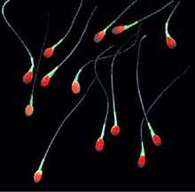
- flagellar motility, a swimming-like motion (observed for example in spermatozoa, propelled by the regular beat of their flagellum, or E. coli, which swims by rotating a helical prokaryotic flagellum)
- amoeboid movement, a crawling-like movement, which also makes swimming possible[13][14]
- gliding motility
- swarming motility
Many cells are not motile, for example Klebsiella pneumoniae and Shigella, or under specific circumstances such as Yersinia pestis at 37 °C.
Muscle Contraction
The nervous system and musculoskeletal system control the majority of mammalian motility. |Gastrointestinal motility is essential for digestion.
Movements
The events that are perceived as movements can be directed:
- along a chemical gradient (see chemotaxis)
- along a temperature gradient (see thermotaxis)
- along a light gradient (see phototaxis)
- along a magnetic field line (see magnetotaxis)
- along an electric field (see galvanotaxis)
- along the direction of the gravitational force (see gravitaxis)
- along a rigidity gradient (see durotaxis)
- along a gradient of cell adhesion sites (see haptotaxis)
- along other cells or biopolymers
 Our muscles give us the ability to move voluntarily (e.g. to throw a ball) and involuntarily (e.g. muscle spasms and reflexes). At the level of the muscular system, motility is a synonym for locomotion.[15][16]
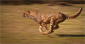
 The record speeds Cheetahs hold are owed in large to their muscle motility.
 The shoots of plants move by growing towards light. This is known as positive phototropism. The roots grow away from light. This is known as negative phototropism.
 Monocytes and macrophages of the immune system engulf Bacteria by extending their pseudopodia. Note that this cartoon is not an accurate representation of phagocytosis.
 Motility becomes very complicated at the sub-cellular level. Shown here is a simplified video animation of translation - a highly motile molecular process.
See also
References
- ↑ Clegg, Chris (2008). "3.2 Cells make organisms". Edexcel biology for AS (6th ed.). London: Hodder Murray. p. 111. ISBN 978-0-340-96623-5.
Division of the cytoplasm, known as cytokinesis, follows telophase. During division, cell organelles such as mitochondria and chloroplasts become distributed evenly between the cells. In animal cells, division is by in-tucking of the plasma membrane at the equator of the spindle, 'pinching' the cytoplasm in half (Figure 3.15). In plant cells, the Golgi apparatus forms vesicles of new cell wall materials which collect along the line of the equator of the spindle, known as the cell plate. Here, the vesicles coalesce forming the new plasma membranes and cell walls between the two cells (Figure 3.17).
- ↑ Alberts, Bruce; Johnson, Alexander; Lewis, Juian; Raff, Martin; Roberts, Keith; Walter, Peter (2008). "16". Molecular biology of the cell (5th ed.). New York: Garland Science. p. 965. ISBN 0-8153-4106-7.
For cells to function properly, they must organize themselves in space and interact mechanically with their environment... Eucaryotic cells have developed... the cytoskeleton... pulls the chromosomes apart at mitosis and then splits the dividing cell into two... drives and guides intracellular traffic of organelles... enables cells such as sperm to swim and others, such as fibroblasts and white blood cells, to crawl across surfaces.
- ↑ "Online Etymology Dictionary".
"capacity of movement," 1827, from French motilité (1827), from Latin mot-, stem of movere "to move" (see move (v.)).
- ↑ Nüsslein-Volhard, Christiane (2006). "6 Form and Form Changes". Coming to life: how genes drive development. [San Diego, CA]: Kales Press. p. 75. ISBN 0979845602.
During development, any change in cell shape is preceded by a change in gene activity. It is the cell's origin and environment that determine which transcription factors are active within a cell, and, hence, which genes are turned on, and which proteins are produced.
- ↑ Fullick, Ann (2009). "7.1". Edexcel A2-level biology. Harlow: Pearson. p. 138. ISBN 978-1-4082-0602-7.
Cyanide is well known in murder mysteries - and has been used in real murders too. The poison acts on cytochrome oxidase in the electron transport chain, preventing the production of ATP. The cells of the body cannot function without their energy supply, so the muscles spasm and the victim cannot breathe.
- ↑ Fullick, Ann (2009). "6.1". Edexcel A2-level biology. Harlow: Pearson. p. 67. ISBN 978-1-4082-0602-7.
As the muscles run out of ATP, the muscle fibres become permanently contracted and lock solid. This produces a stiffening effect which is known as rigor mortis.
- ↑ E. Cooper, Chris; C. Brown, Guy (October 2008). "The inhibition of mitochondrial cytochrome oxidase by the gases carbon monoxide, nitric oxide, hydrogen cyanide and hydrogen sulfide: chemical mechanism and physiological significance". Journal of Bioenergetics and Biomembranes. 40 (5): 533–539. doi:10.1007/s10863-008-9166-6.
- ↑ Krohn, Martha M.; Boisdair, Daniel (May 1994). "Use of a Stereo-video System to Estimate the Energy Expenditure of Free-swimming Fish". Canadian Journal of Fisheries and Aquatic Sciences. 51 (5): 1119–1127. doi:10.1139/f94-111.
- ↑ Cooke, Steven J.; Thorstad, Eva B.; Hinch, Scott G. (March 2004). "Activity and energetics of free-swimming fish: insights from electromyogram telemetry". Fish and Fisheries. 5 (1): 21–52. doi:10.1111/j.1467-2960.2004.00136.x.
We encourage the continued development and refinement of devices for monitoring the activity and energetics of free-swimming fish
- ↑ Carey, Francis G.; Lawson, Kenneth D. (February 1973). "Temperature regulation in free-swimming bluefin tuna". Comparative Biochemistry and Physiology A. 44 (2): 375–392. doi:10.1016/0300-9629(73)90490-8.
Acoustic telemetry was used to monitor ambient water temperature and tissue temperature in free-swimming bluefin tuna (Thunnus thynnus Linneaus [sic], 1758) over periods ranging from a few hours to several days.
- ↑ Intestinal Motility Disorders at eMedicine
- ↑ Wildmarier, Eric P.; Raff, Hershel; Strang, Kevin T. (2016). Vander's Human Physiology: The Mechanisms of Body Function (14th ed). New York, NY: McGraw Hill. p. 528.
- ↑ Van Haastert, Peter J. M. (2011). "Amoeboid Cells Use Protrusions for Walking, Gliding and Swimming". PLoS ONE. 6 (11): e27532. Bibcode:2011PLoSO...627532V. PMC 3212573
 . PMID 22096590. doi:10.1371/journal.pone.0027532.
 - ↑ Bae, A. J.; Bodenschatz, E. (2010). "On the swimming of Dictyostelium amoebae". Proceedings of the National Academy of Sciences. 107 (44): E165–6. Bibcode:2010PNAS..107E.165B. PMC 2973909
 . PMID 20921382. doi:10.1073/pnas.1011900107.
 - ↑ Parsons, Richard (2009). "Unit 5 Section 1". A2-level biology : the revision guide : exam board: Edexcel. Broughton-in-Furness: Coordination Group Publications. p. 50. ISBN 978-1-84762-264-8.
Skeletal muscle is the type of muscle you use to move, e.g. the bicep and triceps move the lower arm. Skeletal muscles are attached to bones by tendons. Ligaments attach bones to other bones, to hold them together. Skeletal muscles contract and relax to move bones at a joint.
- ↑ Vannini, Vanio; Jolly, Richard T.; Pogliani, Giuliano (1994). The new atlas of the human body : a full color guide to the structure of the body. London: Chancellor Press. p. 25. ISBN 1-85152-984-5.
The muscle mass is not just concerned with locomotion. It assists in the circulation of blood and protects and confines the visceral organs. It also provides the main shaping component of the human form.