Middle cerebellar peduncle
| Middle cerebellar peduncle | |
|---|---|
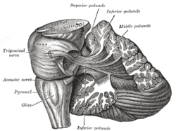
 Dissection showing the projection fibers of the cerebellum. (Middle peduncle labeled at upper right.) | |
| Details | |
| Identifiers | |
| Latin | pedunculus cerebellaris medius |
| NeuroNames | hier-616 |
| NeuroLex ID | Middle cerebellar peduncles |
| Dorlands /Elsevier | p_10/12622515 |
| TA |
A14.1.05.003 A14.1.07.416 |
| FMA | 72515 |
The middle cerebellar peduncles (brachia pontis) are paired structures (left and right) that connect the cerebellum to the pons and are composed entirely of centripetal fibers, which arise from the pontine nucleus of the opposite hemisphere of the cerebellar cortex. The fibers are arranged in three fasciculi: superior, inferior, and deep.
- The superior fasciculus, the most superficial, is derived from the upper transverse fibers of the pons; it is directed backward and lateralward superficial to the other two fasciculi, and is distributed mainly to the lobules on the inferior surface of the cerebellar hemisphere and to the parts of the superior surface adjoining the posterior and lateral margins.
- The inferior fasciculus is formed by the lowest transverse fibers of the pons; it passes under cover of the superior fasciculus and is continued downward and backward more or less parallel with it, to be distributed to the folia on the under surface close to the vermis.
- The deep fasciculus comprises most of the deep transverse fibers of the pons. It is at first covered by the superior and inferior fasciculi, but crosses obliquely and appears on the medial side of the superior, from which it receives a bundle; its fibers spread out and pass to the upper anterior cerebellar folia. The fibers of this fasciculus cover those of the inferior cerebellar peduncle.
Additional images
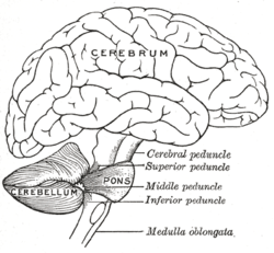
 Scheme showing the connections of the several parts of the brain.
 Superficial dissection of brain-stem. Lateral view.
 Hind- and mid-brains; postero-lateral view.
 Upper part of medulla spinalis and hind- and mid-brains; posterior aspect, exposed in situ.
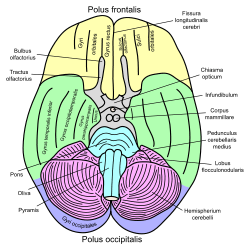
 Basal view of a human brain
- Dissection of human midbrain with middle cerebellar peduncle labeled.
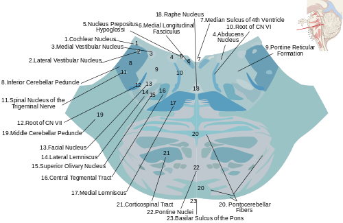
 Cross section through lower pons showing part of the middle cerebellar peduncle (#19) forming from the convergence of pontocerebellar fibers.
- Middle cerebellar peduncle
- Cerebrum. Deep dissection. Inferior dissection.
- Fourth ventricle. Posterior view.Deep dissection.
- Cerebrum.Inferior view.Deep dissection.
- Cerebrum.Inferior view.Deep dissection.
- Cerebrum.Inferior view.Deep dissection.
- Cerebellum. Inferior surface.
- Cerebellum. Inferior surface.
- Cerebellum. Inferior surface.
References
This article incorporates text in the public domain from the 20th edition of Gray's Anatomy (1918)
External links
- Atlas image: n2a7p4 at the University of Michigan Health System
This article is issued from
Wikipedia.
The text is licensed under Creative Commons - Attribution - Sharealike.
Additional terms may apply for the media files.