Lingulodinium polyedrum
| Lingulodinium polyedrum | |
|---|---|
 | |
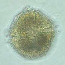
| Ventral view of Lingulodinium polyedrum 900x magnification | |
| Scientific classification | |
| Domain: | Eukaryota |
| (unranked): | SAR |
| (unranked): | Alveolata |
| Phylum: | Dinoflagellata |
| Class: | Dinophyceae |
| Order: | Gonyaulacales |
| Family: | Gonyaulacaceae |
| Genus: | Lingulodinium |
| Species: | L. polyedrum |
| Binomial name | |
| Lingulodinium polyedrum Dodge, 1989 | |
Lingulodinium polyedrum is a species of motile dinoflagellates (synonym Gonyaulax polyedra), which produces a dinoflagellate cyst called Lingulodinium machaerophorum (synonym Hystrichosphaeridium machaerophorum).
Life cycle
As part of its life cycle, this species produces a resting stage, a dinoflagellate cyst called Lingulodinium machaerophorum (synonym Hystrichosphaeridium machaerophorum). This cyst was first described by Deflandre and Cookson in 1955 from the Miocene of Balcombe Bay, Victoria, Australia as: "Shell globular, subsphaerical or ellipsoidal with a rigid membrane, more brittle than deformable, covered with numerous long, stiff, conical, pointed processes resembling the blade of a dagger. Surface of shell granular or punctate."[1] Its stratigraphic range is the Upper Paleocene of eastern USA [2] and Denmark [3] till Recent.
Organic-walled dinocyst morphology is shown to be controlled by changes in salinity and temperature in some species, more particularly process length variation (processes are sometimes called spines, but that is incorrect because they are not necessarily pointy). This morphological variation is known for Lingulodinium machaerophorum from culture experiments,[4] and study of surface sediments.[5] The morphological variation of process lengths can be applied for the reconstruction of salinity. Process length variation of Lingulodinium machaerophorum has been used to reconstruct Black Sea salinity variation.[6]
Toxicity

Lingulodinium polyedrum has been related to production of Yessotoxins (YTXs), a group of structurally related polyether toxins, which can accumulate in shellfish and can produce symptoms similar to those produced by Paralytic Shellfish Poisoning (PSP) toxins.[7]
Luminescence
Lingulodinium polyedrum produces brilliant displays of bioluminescence in warm coastal waters. Seen in Southern California regularly since at least 1901.
They are easily visible under 100x magnification (use the 10x or "scanning" objective on most compound microscopes) and their scintillons luminesce in response to surface tension and acidity. Luminescence is under circadian regulation, peaking at night. Because of this obvious rhythms (and also due to the fact that most its activities, physiological and molecular, are rhythmic) L. polyedrum has been a model organism for studying clocks in single cells.[8]
These blooms contain sufficient concentrations of dinoflagellates that they can provide excellent experimental material for students. A jug of water can be collected from the surf and brought into a completely dark classroom. After a minute or so of complete darkness, the organisms will bioluminesce when the bottle is agitated. Vinegar, baking soda, and vegetable oil can be added in drops to see if they affect the luminescence.
Though brilliant to the naked eye, the bioluminescence is relatively dim for photographic purposes. For example, the photograph at right required a 3 second exposure at f4 at an ISO of 3200, a 210 mm telephoto lens, and the photographer had to hold his hand over the bottom half of the lens to block out the ambient light reflecting off of seafoam in the foreground.
One fantastic experience is that the organisms are in the beach sand too, so when one walks on the beach at night when they are blooming, they luminesce in response to the shock of one's foot hitting the ground.
References
- ↑ Deflandre, G. and Cookson, I.C., 1955. Fossil microplankton from Australian Late Mesozoic and Tertiary sediments. Aust. J. Mar Freshw. Res. 6, 242-313.
- ↑ EDWARDS, L.E., GOODMAN, D.K., and WITMER, R.J. 1984 Lower Tertiary (Pamunkey Group) dinoflagellate biostratigraphy, Potomac River area, Virginia and Maryland. In: Frederiksen, N.O., and Krafft, K. (eds.), Cretaceous and Tertiary stratigraphy, paleontology, and structure, southwestern Maryland and northeastern Virginia—Field trip volume and guide book. American Association of Stratigraphic Palynologists Foundation, Dallas, Texas, p. 137–152.
- ↑ HEILMANN-CLAUSEN, C. 1985 Dinoflagellate stratigraphy of the uppermost Danian to Ypressian in the Viborg 1 borehole, central Jylland, Denmark. Danmarks Geologiske Undersøgelse, Serie A, 7: 1–69.
- ↑ Hallett, R.I., 1999. Consequences of environmental change on the growth and morphology of Lingulodinium polyedrum (Dinophyceae) in culture. Ph.D. thesis. University of Westminster, 109 pp.
- ↑ Mertens, K. N., Ribeiro, S. Bouimetarhan, I., Caner, H., Combourieu-Nebout, N. Dale, B., de Vernal, A. Ellegaard, M. Filipova, M., Godhe, A. Grøsfjeld, K. Holzwarth, U. Kotthoff, U. Leroy, S., Londeix, L., Marret, F., Matsuoka, K., Mudie, P., Naudts, L., Peña-manjarrez, J., Persson, A., Popescu, S., Sangiorgi, F., van der Meer, M., Vink, A., Zonneveld, K., Vercauteren, D., Vlassenbroeck, J., Louwye, S., 2009a. Process length variation in cysts of a dinoflagellate, Lingulodinium machaerophorum, in surface sediments investigating its potential as salinity proxy. Marine Micropaleontology 70, 54–69.
- ↑ Mertens, K.N., Bradley, L.R., Takano, Y., Mudie, P.J., Marret, F., Aksu, A.E., Hiscott, R.N., Verleye, T.J., Mousing, E.A., Smyrnova, L.L., Bagheri, S., Mansor, M., Pospelova, V. & Matsuoka, K. 2012. Quantitative estimation of Holocene surface salinity variation in the Black Sea using dinoflagellate cyst process length. Quaternary Science Reviews.
- ↑ Paz et al. 2008. Yessotoxins, a Group of Marine Polyether Toxins: an Overview. Mar. Drugs 2008, 6, 73-102; DOI: 10.3390/md20080005
- ↑ Hastings JW. 2008. The Gonyaulax clock at 50: translational control of circadian expression. Cold Spring Harb Symp Quant Biol. 2007;72:141-4. doi: 10.1101/sqb.2007.72.026
Further reading
Riccardi, M; Guerrini, F; Ventrella, Ventrella (May 2010). "Lipid and DNA features of Gonyaulax fragilis (Dinophyceae) as potential biomarkers in mucilage genesis". HARMFUL ALGAE. 9 (4): 359–366. doi:10.1016/j.hal.2010.01.004.
External links
- North County Times interview of Dr Franks regarding L polyedrum
- biochemistry of scintillons
- UC Santa Cruz Phytoplankton Identification page