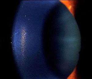
Krukenberg's spindle
| Krukenberg's Spindle | |
|---|---|
 | |
| Slit lamp photograph showing Krukenberg's Spindle as pigment cell deposits on the cornea | |
| Classification and external resources | |
| ICD-9-CM | 364.53 |
| OMIM | 600510 |
| DiseasesDB | 31301 |
Krukenberg's spindle is the name given to the pattern formed on the inner surface of the cornea by pigmented iris cells which are deposited as a result of the currents of the aqueous humor. The sign was described in 1899 by Friedrich Ernst Krukenberg (1871-1946), who was a German pathologist specialising in Ophthalmology.[1]
Differential diagnosis
Iritis
- Painful red eye with photophobia associated with inflammation
Vortex keratopathy
- Corneal deposits also known as Cornea verticillata, caused by chronic amiodarone use for cardiac arrhythmias.[2]
Corneal guttata
- Non-transparent collagen deposits appearing following loss of corneal endothelial cells[3]
See also
References
- ↑ Krukenberg F. (1899) Beiderseitige angeborene Melanose der Hornhaut. Klin Mbl Augenheilkd 37:254-258.
- ↑ Chew, E; Ghosh, M; McCulloch, C (June 1982). "Amiodarone-induced cornea verticillata.". Canadian journal of ophthalmology. Journal canadien d'ophtalmologie. 17 (3): 96–9. PMID 7116220.
- ↑ Akimune C, Watanabe H, Maeda N, et al. (January 2000). "Corneal guttata associated with the corneal dystrophy resulting from a betaig-h3 R124H mutation". 84 (1): 67–71. PMC 1723238
 . PMID 10611102.
This article is issued from
Wikipedia.
The text is licensed under Creative Commons - Attribution - Sharealike.
Additional terms may apply for the media files.