Ki-67 (protein)

Antigen KI-67 also known as Ki-67 or MKI67 is a protein that in humans is encoded by the MKI67 gene (antigen identified by monoclonal antibody Ki-67).[3][4][5]
Function
Antigen KI-67 is a nuclear protein that is associated with and may be necessary for cellular proliferation. Furthermore, it is associated with ribosomal RNA transcription.[5] Inactivation of antigen KI-67 leads to inhibition of ribosomal RNA synthesis.[6]
Use as a marker of proliferating cells

The Ki-67 protein (also known as MKI67) is a cellular marker for proliferation.[7] It is strictly associated with cell proliferation. During interphase, the Ki-67 antigen can be exclusively detected within the cell nucleus, whereas in mitosis most of the protein is relocated to the surface of the chromosomes.[8] Ki-67 protein is present during all active phases of the cell cycle (G1, S, G2, and mitosis), but is absent in resting (quiescent) cells (G0).[9] Cellular content of Ki-67 protein markedly increases during cell progression through S phase of the cell cycle.[10]
Antibody labeling
Ki-67 is an excellent marker to determine the growth fraction of a given cell population. The fraction of Ki-67-positive tumor cells (the Ki-67 labeling index) is often correlated with the clinical course of cancer. The best-studied examples in this context are carcinomas of the prostate, brain and the breast and nephroblastoma. For these types of tumors, the prognostic value for survival and tumor recurrence have repeatedly been proven in uni- and multivariate analysis.
MIB-1
Ki-67 and MIB-1 monoclonal antibodies are directed against different epitopes of the same proliferation-related antigen. Ki-67 and MIB1 may be used on fixed sections.[11] MIB-1 is used in clinical applications to determine the Ki-67 labelling index. One of its primary advantages over the original Ki-67 antibody (and the reason why it has essentially supplanted the original antibody for clinical use) is that it can be used on formalin-fixed paraffin-embedded sections, after heat-mediated antigen retrieval (see next section below).
Original Ki-67 antibody
The Ki-67 protein was originally defined by the prototype monoclonal antibody Ki-67,[12] which was generated by immunizing mice with nuclei of the Hodgkin lymphoma cell line L428. The name is derived from the city of origin (Kiel, Germany) and the number of the original clone in the 96-well plate.
Interactions
Ki-67 (protein) has been shown to interact with CBX3.[13]
See also
- PCNA - Proliferating Cell Nuclear Antigen, expressed during the DNA synthesis.
Additional images
 Immunofluorescent antibody staining against neurofilament (green) and Ki-67 (red) in a mouse embryo 12.5 days after fertilization. The proliferating cells are in the ventricular zone in the neural tube and therefore colored red.
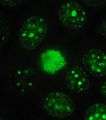 Protein Ki-67 in human MCF-7 cells.
 Ki-67 protein (red), tubulin (green) and DNA (blue) in HeLa cells. Dividing cells show strong Ki-67 staining in cell nuclei while all cells contain large amounts of tubulin, the major component of microtubules. Antibodies, cell staining and image courtesy of EnCor Biotechnology.
References
- ↑ "Human PubMed Reference:".
- ↑ "Mouse PubMed Reference:".
- ↑ "Entrez Gene: Antigen identified by monoclonal antibody Ki-67".
- ↑ Schonk DM, Kuijpers HJ, van Drunen E, van Dalen CH, Geurts van Kessel AH, Verheijen R, Ramaekers FC (October 1989). "Assignment of the gene(s) involved in the expression of the proliferation-related Ki-67 antigen to human chromosome 10". Hum. Genet. 83 (3): 297–9. PMID 2571566. doi:10.1007/BF00285178.
- 1 2 Bullwinkel J, Baron-Lühr B, Lüdemann A, Wohlenberg C, Gerdes J, Scholzen T (March 2006). "Ki-67 protein is associated with ribosomal RNA transcription in quiescent and proliferating cells". J. Cell. Physiol. 206 (3): 624–35. PMID 16206250. doi:10.1002/jcp.20494.
- ↑ Rahmanzadeh R, Hüttmann G, Gerdes J, Scholzen T (June 2007). "Chromophore-assisted light inactivation of pKi-67 leads to inhibition of ribosomal RNA synthesis". Cell Prolif. 40 (3): 422–30. PMID 17531085. doi:10.1111/j.1365-2184.2007.00433.x.
- ↑ Scholzen T, Gerdes J (March 2000). "The Ki-67 protein: from the known and the unknown". Journal of Cellular Physiology. 182 (3): 311–22. PMID 10653597. doi:10.1002/(SICI)1097-4652(200003)182:3<311::AID-JCP1>3.0.CO;2-9.
- ↑ Cuylen S, Blaukopf C, Politi AZ, Müller-Reichert T, Neumann B, Poser I, Ellenberg J, Hyman AA, Gerlich DW (July 2016). "Ki-67 acts as a biological surfactant to disperse mitotic chromosomes". Nature. 535 (7611): 308–12. PMID 27362226. doi:10.1038/nature18610.
- ↑ Bruno S, Darzynkiewicz Z (January 1992). "Cell cycle dependent expression and stability of the nuclear protein detected by Ki-67 antibody in HL-60 cells". Cell Proliferation. 25 (1): 31–40. PMID 1540682. doi:10.1111/j.1365-2184.1992.tb01435.x.
- ↑ Darzynkiewicz Z, Zhao H, Zhang S, Lee MY, Lee EY, Zhang Z (May 2015). "Initiation and termination of DNA replication during S phase in relation to cyclins D1, E and A, p21WAF1, Cdt1 and the p12 subunit of DNA polymerase δ revealed in individual cells by cytometry". Oncotarget. 6 (14): 11735–50. PMID 26059433. doi:10.18632/oncotarget.4149.
- ↑ Bánkfalvi A (November 2000). "Comparative methodological analysis of erbB-2/HER-2 gene dosage, chromosomal copy number and protein overexpression in breast carcinoma tissues for diagnostic use.". Histopathology. 37 (5): 411–9. PMID 11119122. doi:10.1046/j.1365-2559.2000.00984.x.
- ↑ Gerdes J, Schwab U, Lemke H, Stein H (1983). "Production of a mouse monoclonal antibody reactive with a human nuclear antigen associated with cell proliferation". Int. J. Cancer. 31 (1): 13–20. PMID 6339421. doi:10.1002/ijc.2910310104.
- ↑ Kametaka, Ai; Takagi Masatoshi; Hayakawa Tomohiro; Haraguchi Tokuko; Hiraoka Yasushi; Yoneda Yoshihiro (Dec 2002). "Interaction of the chromatin compaction-inducing domain (LR domain) of Ki-67 antigen with HP1 proteins". Genes Cells. England. 7 (12): 1231–42. ISSN 1356-9597. PMID 12485163. doi:10.1046/j.1365-2443.2002.00596.x.
External links
- Ki-67 Antigen at the US National Library of Medicine Medical Subject Headings (MeSH)
- http://www.pathologyoutlines.com/topic/stainski67.html
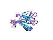
PDB gallery | ||||||
|---|---|---|---|---|---|---|
| ||||||