Bulbourethral gland
| Bulbourethral gland | |
|---|---|
|
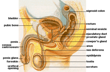
Male Anatomy | |
|
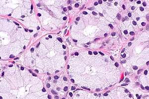
Micrograph of bulbourethral gland. H&E stain. | |
| Details | |
| Precursor | Urogenital sinus |
| Artery | Artery of the urethral bulb |
| Identifiers | |
| Latin | Glandulae bulbourethrales |
| MeSH | A05.360.444.123 |
A bulbourethral gland, also called a Cowper's gland for English anatomist William Cowper, is one of two small exocrine glands in the reproductive system of many male mammals (of all domesticated animals, they are only absent in the dog).[1] They are homologous to Bartholin's glands in females.
Location
Bulbourethral glands are located posterior and lateral to the membranous portion of the urethra at the base of the penis, between the two layers of the fascia of the urogenital diaphragm, in the deep perineal pouch. They are enclosed by transverse fibers of the sphincter urethrae membranaceae muscle.
Structure
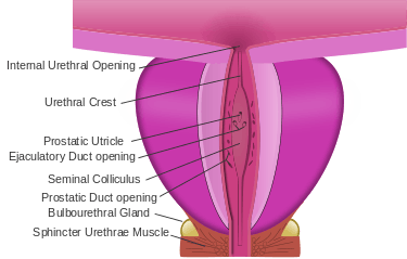
The bulbourethral glands are compound tubulo-alveolar glands, each approximately the size of a pea in humans. In chimpanzees, they are not visible during dissection, but can be found on microscopic examination.[2] In boars, they are up to 18 cm long and 5 cm in diameter.[1] They are composed of several lobules held together by a fibrous covering. Each lobule consists of a number of acini, lined by columnar epithelial cells, opening into a duct that joins with the ducts of other lobules to form a single excretory duct. This duct is approximately 2.5 cm long and opens into the bulbar urethra at the base of the penis. The glands gradually diminish in size with advancing age.[3]
Some human males may excrete sperm carried out of the body by Cowper's gland secretions prior to ejaculation, at concentrations similar to those found in their semen.[4] This could result in the possibility of conception from the introduction of pre-ejaculate alone to the vagina, though the direct probability of pregnancy has not been assessed.
Function
The bulbourethral gland contributes about 0.1 to 0.2 ml or 5% of the ejaculate. The secretion is a clear fluid that is rich in mucoproteins. The secretion also helps to lubricate the distal urethra, and neutralize acidic urine which remains in the urethra.
Gallery
 Structure of the penis
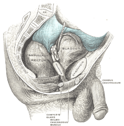 Male pelvic organs seen from right side.
 Vertical section of bladder, penis, and urethra.
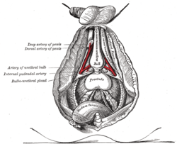 Bulbourethral gland labeled at center left.
See also
References
- 1 2 Mark McEntee (December 2, 2012). Reproductive Pathology of Domestic Mammals. Elsevier Science. p. 333. ISBN 978-0-323-13804-8. Retrieved August 20, 2013.
- ↑ Jeffrey H. Schwartz (1988). Orang-utan Biology. Oxford University Press. p. 92. ISBN 978-0-19-504371-6. Retrieved August 20, 2013.
- ↑ Gray's Anatomy, 38th ed., p 1861.
- ↑ Stephen R. Killick; Christine Leary; James Trussell; Katherine A. Guthrie (March 2011). "Sperm content of pre-ejaculatory fluid". Human Fertility. Informa. 14 (1): 48–52. ISSN 1464-7273. PMC 3564677
 . PMID 21155689. doi:10.3109/14647273.2010.520798.
