Common cardinal veins
| Common cardinal veins | |
|---|---|
 Scheme of arrangement of parietal veins. | |
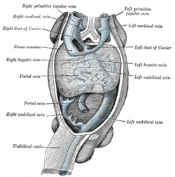
 Human embryo with heart and anterior body-wall removed to show the sinus venosus and its tributaries. | |
| Details | |
| Latin | vena cardinalis communis |
| Dorlands /Elsevier | v_04/12847349 |
During development of the veins, the first indication of a parietal system consists in the appearance of two short transverse veins, the ducts of Cuvier (or common cardinal veins[1]), which open, one on either side, into the sinus venosus. Each of these ducts receives an ascending and descending vein. The ascending veins return the blood from the parietes of the trunk and from the Wolffian bodies, and are called cardinal veins.
Additional images
 Figure obtained by combining several successive sections of a human embryo of about the fourth week.
 Upper part of celom of human embryo of 6.8 mm., seen from behind.
 Dorsal surface of heart of human embryo of thirty-five days.
See also
References
This article incorporates text in the public domain from the 20th edition of Gray's Anatomy (1918)
- ↑ ZFIN: Anatomical Structure: common cardinal vein Archived 2007-09-28 at the Wayback Machine.
External links
- cardev-009—Embryo Images at University of North Carolina
This article is issued from
Wikipedia.
The text is licensed under Creative Commons - Attribution - Sharealike.
Additional terms may apply for the media files.