Cerebrovascular disease
| Cerebrovascular disease | |
|---|---|
 | |
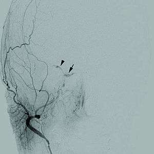
| Cerebral angiogram of a carotid-cavernous fistula | |
| Classification and external resources | |
| Specialty | cardiology, neurology |
| ICD-10 | I60-I69 |
| MeSH | D002561 |
A cerebrovascular disease is a vascular disease of the cerebral circulation. Arteries supplying oxygen to the brain are affected resulting in one of a number of cerebrovascular diseases.[1] Most commonly this is a stroke or mini-stroke and sometimes can be a hemorrhagic stroke.[1] Any of these can result in vascular dementia.[2]
Hypertension (high blood pressure) is the most important contributing cause because it damages the blood vessel lining exposing collagen where platelets aggregate to initiate a repair. If maintained, hypertension can change the structure of blood vessels (narrow, deformed).[3]
Blood pressure affects blood flow in narrowed vessels causing ischemic stroke, a rise in blood pressure can cause tearing of vessels leading to intracranial hemorrhage.[4]
A stroke usually presents with an abrupt onset of a neurologic deficit, attributable to a focal vascular lesion.[5] The neurologic symptoms manifest within seconds because neurons lack glycogen, so energy failure is rapid.[6]
Types of stroke
- Ischemic stroke,the most common is caused by a blockage of a blood vessel in the brain, usually caused by thrombosis or emboli from a proximal arterial source or the heart, that leads to the brain being starved of oxygen.[7] The neurologic signs and symptoms must last longer than 24 hours or the brain infarction is demonstrated, mainly by imaging techniques.[8]
- Transient ischemic attack (TIA) also called a mini-stroke. This is a condition in which the blood flow is quickly restored and the brain tissue can fully recover and the symptoms are only transient, leaving no sequelae.[9] In order to diagnose this entity all neurologic signs and symptoms must have been resolved within 24 hrs without evidence of brain infarction on brain imaging.[10]
- Subarachnoid haemorrhage where blood leaks out of blood vessels directly into or around the brain.[1] The neurologic symptoms are produced by the blood mass effect on neural structures, from the toxic effects of blood on the brain tissue, or by the increasing of intracranial pressure.[11]
Causes
Causes of cerebrovascular disease can be divided into: atherosclerosis, embolism, aneurysms, low flow states, and other rare causes.[12] Major modifiable risk factors include:[13]
Pathophysiology
Once a reduction in blood flow occurs, that lasts seconds the brain tissue suffers ischemia.[14][15] If the interruption of blood flow is not restored in minutes, the tissue suffers infarction followed by tissue death.[16] When the low cerebral blood flow persists for a longer duration, this may develop into an infarction in the border zones (areas of poor blood flow between the major cerebral artery distributions). In more severe instances, global hypoxia-ischemia causes widespread brain injury leading to a severe cognitive sequelae called hypoxic-ischemic encephalopathy.[17]
Strokes can also result from embolisms, furthermore, embolisms block small arteries, causing damage to occur.[4] Spontaneous rupture of a blood vessel in the brain causes a hemorrhagic stroke.[18] Another form of cerebrovascular disease includes aneurysms. Cerebral aneurysms can be genetic in nature, due to a wall deformity of the artery. Such aneurysms are common in individuals with genetic diseases ( connective tissue disorders, polycystic kidney disease, and arteriovenous malformations).[19]
The carotid arteries cover the majority of the cerebrum. The common carotid artery divides into the internal and the external carotid arteries. The internal carotid artery becomes the anterior cerebral artery and the middle central artery. The ACA transmits blood to the frontal parietal. From the basilar artery are two posterior cerebral arteries. Branches of the basilar and PCA supply the occipital lobe, brain stem, and the cerebellum.[20] Ischemia is the loss of blood flow to the focal region of the brain. This produces heterogeneous areas of ischemia at the affected vascular region, furthermore blood flow is limited to a residual flow. Regions with blood flow of less than 10 mL/100 g of tissue/min are core regions (cells here die within minutes of a stroke).The ischemic penumbra with a blood flow of <25 ml/100g tissue/min, remain usable for more time (hours).[21]
An ischemic cascade occurs where an energetic molecular problem arises. ATP consumption continues in spite of insufficient production, this causes total levels of adenosine triphosphate to decrease and lactate acidosis to become established (ionic homeostasis in neurons is lost). The downstream mechanisms of the ischemic cascade thus begins. Ion pumps no longer transport Ca2+ out of cell, this triggers release of glutamate, which in turn allows calcium into cell walls. In the end the apoptosis pathway is initiated and cell death occurs.[22]
Evaluation
Diagnosis of cerebrovascular disease is done by (among other diagnoses):[23]
- clinical history
- physical exam
- neurological examination.
It is important to differentiate the symptoms caused by a stroke from those caused by syncope (fainting) which is also a reduction in cerebral blood flow, almost always generalized, but they are usually caused by systemic hypotension of various origins: cardiac arrhythmias, myocardial infarction, hemorrhagic shock, among others.[24]
Treatment
Treatment for cerebrovascular disease includes medication, lifestyle changes and surgery.[4]
Examples of medications are:
- antiplatelets (aspirin, clopidogrel)
- blood thinners (heparin, warfarin)
- antihypertensives (ACE inhibitors, beta blockers)
- anti-diabetic medications.
Surgical procedures include:
- endovascular surgery and vascular surgery (for future stroke prevention).
Epidemiology

Worldwide, it is estimated there are 31 million stroke survivors, though about 6 million deaths were due to cerebrovascular disease (2nd most common cause of death in the world and 6th most common cause of disability).[26]
Cerebrovascular disease primarily occurs with advanced age; the risk for developing it goes up significantly after 65 years of age. CVD tends to occur earlier than Alzheimer's Disease (which is rare before the age of 80). In some countries such as Japan, CVD is more common than AD.
In 2012 6.4 million US individuals (adults) had a stroke, which corresponds to 2.7% in the U.S. With approximately 129,000 deaths in 2013 (U.S.)[27]
Geographically, a "stroke belt" in the US has long been known, similar to the "diabetes belt"which includes all of Mississippi and parts of Alabama, Arkansas, Florida, Georgia, Kentucky, Louisiana, North Carolina, Ohio, Pennsylvania, South Carolina, Tennessee, Texas, Virginia, and West Virginia.[28]
See also
References
- 1 2 3 "Cerebrovascular disease - Introduction - NHS Choices". www.nhs.uk. Retrieved 2015-09-01.
- ↑ "Vascular dementia - Causes - NHS Choices". www.nhs.uk. Retrieved 2015-09-01.
- ↑ Prakash, Dibya (2014-04-10). Nuclear Medicine: A Guide for Healthcare Professionals and Patients. Springer Science & Business Media. p. 142. ISBN 9788132218265.
- 1 2 3 "Stroke: MedlinePlus Medical Encyclopedia". www.nlm.nih.gov. Retrieved 2015-09-01.
- ↑ "AccessMedicine | Content". accessmedicine.mhmedical.com. Retrieved 2015-11-05.
- ↑ "AccessMedicine | Content". accessmedicine.mhmedical.com. Retrieved 2015-11-05.
- ↑ "Stroke: MedlinePlus". www.nlm.nih.gov. Retrieved 2015-09-01.
- ↑ "Stroke Imaging: Overview, Computed Tomography, Magnetic Resonance Imaging".
- ↑ Sacco, Ralph L.; Kasner, Scott E.; Broderick, Joseph P.; Caplan, Louis R.; Connors, J. J. (Buddy); Culebras, Antonio; Elkind, Mitchell S. V.; George, Mary G.; Hamdan, Allen D. (2013-07-01). "An Updated Definition of Stroke for the 21st Century A Statement for Healthcare Professionals From the American Heart Association/American Stroke Association". Stroke. 44 (7): 2064–2089. ISSN 0039-2499. PMID 23652265. doi:10.1161/STR.0b013e318296aeca.
- ↑ Easton, J. Donald; Saver, Jeffrey L.; Albers, Gregory W.; Alberts, Mark J.; Chaturvedi, Seemant; Feldmann, Edward; Hatsukami, Thomas S.; Higashida, Randall T.; Johnston, S. Claiborne (2009-06-01). "Definition and Evaluation of Transient Ischemic Attack A Scientific Statement for Healthcare Professionals From the American Heart Association/American Stroke Association Stroke Council; Council on Cardiovascular Surgery and Anesthesia; Council on Cardiovascular Radiology and Intervention; Council on Cardiovascular Nursing; and the Interdisciplinary Council on Peripheral Vascular Disease: The American Academy of Neurology affirms the value of this statement as an educational tool for neurologists.". Stroke. 40 (6): 2276–2293. ISSN 0039-2499. PMID 19423857. doi:10.1161/STROKEAHA.108.192218.
- ↑ "Mechanisms of brain injury after intracerebral haemorrhage - The Lancet Neurology". www.thelancet.com. Retrieved 2015-11-05.
- ↑ Corporation, Surgisphere. Clinical Review of Surgery | ABSITE Review. Lulu.com. p. 146. ISBN 9780980210347.
- ↑ "Cerebrovascular disease - NHS Choices - Risks and prevention". www.nhs.uk. Retrieved 2015-09-01.
- ↑ "Cerebral Ischemia | Columbia Neurosurgery". www.columbianeurosurgery.org. Retrieved 2015-11-05.
- ↑ "Stroke: Hope Through Research". www.ninds.nih.gov. Retrieved 2015-11-05.
- ↑ Wang, Hai-rong; Chen, Miao; Wang, Fei-long; Dai, Li-hua; Fei, Ai-hua; Liu, Jia-fu; Li, Hao-jun; Shen, Sa; Liu, Ming (2015-07-24). "Comparison of Therapeutic Effect of Recombinant Tissue Plasminogen Activator by Treatment Time after Onset of Acute Ischemic Stroke". Scientific Reports. 5. PMC 4513278
 . PMID 26206308. doi:10.1038/srep11743.
 - ↑ "Neurological disorders: public health challenges." (PDF). 2006.
- ↑ Herman, Irving P. (2007-02-16). Physics of the Human Body. Springer Science & Business Media. p. 488. ISBN 9783540296041.
- ↑ "Cerebral Aneurysms Fact Sheet: National Institute of Neurological Disorders and Stroke (NINDS)". www.ninds.nih.gov. Retrieved 2015-09-02.
- ↑ Cipolla, Marilyn J. (2009-01-01). "Anatomy and Ultrastructure".
- ↑ "Ischemic Stroke: Practice Essentials, Background, Anatomy".
- ↑ Xing, Changhong; Arai, Ken; Lo, Eng H.; Hommel, Marc (2012-07-01). "Pathophysiologic cascades in ischemic stroke". International Journal of Stroke. 7 (5): 378–385. ISSN 1747-4930. PMC 3985770
 . PMID 22712739. doi:10.1111/j.1747-4949.2012.00839.x.
 - ↑ "Stroke. Diagnosis and Therapeutic Management of Cerebrovascular Disease | Revista Española de Cardiología (English Version)". www.revespcardiol.org. Retrieved 2015-09-01.
- ↑ Bonanno, Fabrizio Giuseppe (2011-01-01). "Clinical pathology of the shock syndromes". Journal of Emergencies, Trauma, and Shock. 4 (2): 233–243. ISSN 0974-2700. PMC 3132364
 . PMID 21769211. doi:10.4103/0974-2700.82211.
 - ↑ "WHO Disease and injury country estimates". World Health Organization. 2009. Retrieved Nov 11, 2009.
- ↑ Ward, Helen; Toledano, Mireille B.; Shaddick, Gavin; Davies, Bethan; Elliott, Paul (2012-05-24). Oxford Handbook of Epidemiology for Clinicians. OUP Oxford. p. 310. ISBN 9780191654787.
- ↑ "FastStats". www.cdc.gov. Retrieved 2015-09-01.
- ↑ Borhani NO. Changes and geographic distribution of mortality from cerebrovascular disease. Am J Public Health Nations Health 1965;55:673–81.
Further reading
- Chan, Pak H. (2002-03-28). Cerebrovascular Disease: 22nd Princeton Conference. Cambridge University Press. ISBN 9781139439657.
- Mark, Steven D.; Wang, Wen; Fraumeni, Joseph F.; Li, Jun-Yao; Taylor, Philip R.; Wang, Guo-Qing; Guo, Wande; Dawsey, Sanford M.; Li, Bing (1996-04-01). "Lowered Risks of Hypertension and Cerebrovascular Disease after Vitamin/Mineral Supplementation The Linxian Nutrition Intervention Trial". American Journal of Epidemiology. 143 (7): 658–664. ISSN 0002-9262. PMID 8651227. doi:10.1093/oxfordjournals.aje.a008798.
- Ning, MingMing; Lopez, Mary; Cao, Jing; Buonanno, Ferdinando S.; Lo, Eng H. (2012-12-01). "Application of proteomics to cerebrovascular disease". Electrophoresis. 33 (24). ISSN 0173-0835. PMC 3712851
 . PMID 23161401. doi:10.1002/elps.201200481.
| Wikimedia Commons has media related to Cerebrovascular diseases. |
