Distal radius fracture
| Distal radius fracture | |
|---|---|
| Classification and external resources | |
|
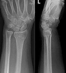
Colles fracture on X-ray | |
| ICD-10 | S52.5 |
| AO | 21-A1 - 21-C3 |
| MeSH | S52.5 |
A distal radius fracture is a common bone fracture of the radius in the forearm. Because of its proximity to the wrist joint, this injury is often called a wrist fracture. Treatment is usually with immobilization, although surgery is sometimes needed for complex fractures.
Specific types of distal radius fractures are Colles' fracture; Smith's fracture; Barton's fracture; Chauffeur's fracture (so called because the crank used to start old cars often kicked back and broke the chauffeurs' wrists with a particular pattern). Most of these names are applied to specific patterns of distal radius fracture but confusion exists because "Colles' fracture" is used (for example by the US National Library of Medicine) as a generic term for distal radius fracture.[1]
Signs and symptoms
History
The most common cause of this type of fracture is a fall on an outstretched hand (acronym: FOOSH).[2] In young adults this fracture is the result of moderate to severe force such as a fall from a significant height or a motor vehicle accident. The risk of injury is increased in patients with osteoporosis and other metabolic bone diseases.
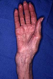
Patients usually present with a history of an injury and localized pain. There is often a deformity in the wrist with associated swelling (figure 1). Numbness of the hand can occur because of compression on the median nerve across the wrist (carpal tunnel syndrome). The wrist deformity often limits motion of the fingers.

Examination
Swelling, deformity, tenderness and loss of wrist motion are normal features on examination of a patient with a distal radius fracture. Examination should rule out a skin wound which might suggest an open fracture. It is imperative to check for loss of sensation, loss of circulation to the hand, and more proximal injuries to the forearm, elbow and shoulder. The most common associated neurological finding is decreased sensation over the thenar eminence due to associated median nerve injury.
Injuries associated
The most commonly associated injury is to the ulnar styloid. Styloid fractures can occur either to the very tip of the styloid or at the base. Because the triangular fibrocartilage (TFCC) attaches to the base of the ulna styloid, displaced fractures can result in instability of the distal radio-ulnar joint. Carpal bone fractures such as those to the scaphoid have been described, whereas instability or dislocations of the wrist are seen with certain types of distal radius and ulna fractures. Injuries to the elbow, humerus and shoulder are also common after a FOOSH (fall on outstretched hand). Swelling and displacement can cause compression on the median nerve across the wrist, an acute carpal tunnel syndrome. Very rarely is pressure on the muscle components of the hand or forearm sufficient to create a compartment syndrome.
Diagnosis
Diagnosis may be evident clinically when the distal radius is deformed but should be confirmed by X-ray. The differential diagnosis includes scaphoid fractures and wrist dislocations, which can also co-exist with a distal radius fracture. Occasionally, fractures may not be seen on X-rays immediately after the injury. Delayed X-rays, X-ray computed tomography (CT scan), or Magnetic resonance imaging (MRI) will confirm the diagnosis.
Medical imaging
X-ray of the affected wrist is required if a fracture is suspected. CT scan is often performed to investigate the articular anatomy of the fracture, especially if surgery is considered. Investigation of a potential distal radial fracture includes assessment of the angle of the joint surface on lateral X-ray, the loss of length of the radius from the collapse of the fracture, and congruency of the distal radioulnar joint. Displacement of the articular surface is the most important factor affecting prognosis and treatment.
Articular surface
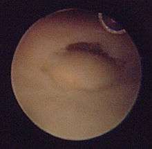
The articular joint's surface must be smooth for it to function properly. Irregularity may result in radiocarpal arthritis, pain, and stiffness. More than 1 mm of incongruity places the patient at a high risk for post-traumatic arthritis. Significant articular incongruity typically occurs in young patients after high energy injuries (Figure 2). If the surface is very irregular and cannot be reconstructed, then the only option may be a fusion.

Lateral articular angle
The lateral articular angle is the angle on an x-ray film between the axis of the radius and the articular cup. Normally, the angle is tilted toward the thumb (volar/ventral tilt) by 11°. The most common fracture pattern usually demonstrates malalignment of this angle and collapse in a dorsal direction. Alignment up to 0° is still considered to be functional, and does not require any intervention. However, tilt away from the thumb (dorsal tilt) beyond this point (>11° deviation) begins to create biomechanical changes that can lead to arthritis, pain, and stiffness. When dorsal tilt beyond the acceptable threshold occurs, distal radio-ulnar joint motion is altered, and forearm rotation becomes restricted. The upper limit of an acceptable deformity after reduction of the fracture is 20° of dorsal tilt.
Radial length
Radial length is an important consideration in distal radius fractures. Distal radius fractures typically result in loss of length as the radius collapses from the loading force of the injury. With increasing relative lengthening of the uninjured ulna, ulnar positive variance, ulnar impaction syndrome may occur. Ulnar impaction syndrome is a painful condition of excessive contact and wear between the ulna and the carpus with an associated is a degenerative tear of the TFCC (Figure 3).

Classification
In medicine, classifications systems are devised to describe patterns of injury which will behave in predictable ways, to distinguish between conditions which have different outcomes or which need different treatments. Most wrist fracture systems have failed to accomplish any of these goals and there is no consensus about the most useful one.
At one extreme, a stable undisplaced extra-articular fracture has an excellent prognosis. On the other hand, an unstable, displaced intra-articular fracture is difficult to treat and has a poor prognosis without operative intervention.
Eponyms such as Colles', Smith's, and Barton's fractures are discouraged. Though the Frykman classification system has traditionally been used, there is little value in its use because it does not help direct treatment. The Universal classification system is descriptive but also does not direct treatment. Universal codes include:
- Type I: extra articular, undisplaced
- Type II: extra articular, displaced
- Type III intra articular, undisplaced
- Type IV: intra articular, displaced
The system that comes closest to directing treatment has been devised by Melone:
- I Stable fracture
- II Unstable "die-punch"
- III "Spike" fracture
- IV Split fracture
- V Explosion injuries
However, an anatomic description of the fracture is the easiest way to describe the fracture, decide on treatment, and make an assessment of stability.
- Articular incongruity
- Radial shortening
- Radial angulation
- Comminution of the fracture (the amount of crumbling at the fracture site)
- Open (compound fracture) or closed injury
- Associated ulnar styloid fracture
- Associated soft tissue injuries
Natural history
Nonunion is rare; almost all of these fractures heal. However, if the fracture is unstable the deformity at the fracture site will increase and cause limitation of wrist motion and forearm rotation, pronation and supination. If the joint surface is damaged and heals with more than 1–2 mm of unevenness, the wrist joint will be prone to post-traumatic osteoarthritis (Figures 4 and 5). Displaced fractures of the ulnar styloid base associated with a distal radius fracture result in instability of the distal radioulnar joint and resulting loss of forearm rotation (Figure 6).
Treatment


The type of treatment required depends on many factors, including displacement and stability of the fracture fragments.
Non-operative
For torus fractures a splint may be sufficient and casting may be avoided.[3]
Where the fracture is undisplaced and stable, non operative treatment involves immobilization. Initially the wrist is splinted to allow swelling and subsequently a cast is applied. Depending on the nature of the fracture, the cast may be placed above the elbow to control forearm rotation.
In displaced fractures, the fracture may be manipulated under anaesthesia and splinted in a position to minimize the risk of re-displacement. Typically, this involves injecting local anesthesia into the fracture (hematoma block) possibly combined with intravenous medication. A manual reduction is performed to reposition the displaced distal radius into its preinjury position and maintain this position in a well formed splint or cast.
During the period of follow-up, it is common practice to repeat x-rays at about 1 week to make sure the position is still acceptable. Further followup is needed to determine when the fracture has healed and when rehabilitation is complete. The critical time during the period of attempted treatment with casting is the first 3 weeks. The swelling will reduce during this time and the fracture can displace. If the displacement becomes unacceptable, closed treatment may need to be abandoned and surgery pursued. More than 3 weeks after injury, the fracture will start to heal and further displacement becomes less likely.
The length of time in the cast varies with different ages. Children heal more rapidly, but may ignore activity restrictions. Three weeks in a cast and 6 weeks off sports may be appropriate for certain fractures. In adults, the risk of stiffness of the joint increases the longer it is immobilized. If callus is seen on x-ray at 4 weeks, the cast may be replaced by a removable splint. However, many hand surgeons leave the patients in the cast for up to 6 weeks. In general, the x-rays will not show any callus until about a month after the fracture is healed; therefore the cast is removed before the x-rays confirm that it is healed.
Displaced fractures in the elderly or those physiologically unable to undergo surgery are treated differently. When the fracture is displaced and there are no plans for a surgery, a short arm cast is placed for only 4 weeks or until the tenderness resolves. A larger cast placed for an extended period of time only slows down recovery in this group of patients.
Following healing and cast removal a period of rehabilitation for recovery of strength and range of motion is necessary. Patients will continue to improve after the fracture for 4 to 12 months.
Reduction
Closed management of a distal radius fracture involves first anesthetizing the affected area with a hematoma block, regional anesthesia, sedation or a general anesthetic.
Manipulation generally includes first placing the arm under traction and unlocking the fragments. The deformity is then reduced with appropriate closed manipulations (depending on the type of deformity) reduction, after which a splint or cast is placed and an X-ray is taken to ensure that the reduction was successful. The cast is usually maintained for about 6 weeks.
Closed treatment is frequently unsuccessful in maintaining a good position in adults, because there is frequently comminution of the fracture. Re-displacement and deformity can reoccur with an unacceptable ultimate result.
Risks of non-operative treatment
Failure of non-operative treatment is common and is the largest risk of an adverse outcome. Studies have shown that the fracture often re-displaces to its original position even in a cast.[4] Only 27% - 32% of fractures are in acceptable alignment 5 weeks after closed reduction.[5] In the long term this increases the risk of stiffness and post traumatic osteoarthritis leading to wrist pain and loss of function. It is because of these findings that many surgeons recommend operative intervention if the fracture is displaced enough to consider a reduction. Ultimately, the fractures that have a closed reduction may return to the position before the reduction is attempted.
Stiffness of the wrist is universal following a fracture of the distal radius. The degree of stiffness in the wrist is dependent on the type of fracture and the period of immobilization. It is for this reason that an open reduction is advantageous. It is also quite common for patients to develop digital stiffness after a fracture of the distal radius. Aggressive movement of the digits while immobilized or following operative treatment is stressed to minimize digital stiffness.
Other risks specific to cast treatment relate to the potential for compression of the swollen arm causing carpal tunnel syndrome or compartment syndrome. Carpal tunnel syndrome may be related to the position of the wrist (i.e. excessive flexion) or excess distraction if the wrist is placed in an external fixator. Compartment syndrome is swelling in the muscle compartments, usually in the forearm, leading to severe pain, loss of nerve function and a contracture. Finally, complex regional pain syndrome (reflex sympathetic dystrophy) is a serious complication following injury and is thought to be more common after cast immobilization than after surgery. The provoking factors for regional pain syndromes, however, are very complex but the condition often leads to chronic pain and stiffness.
Prognosis following non-operative treatment
In children the outcome of distal radius fracture treatment in casts is usually very successful with healing and return to normal function expected. Some residual deformity is common but this often remodels as the child grows. In the elderly, distal radius fractures heal and may result in adequate function following non-operative treatment. A large proportion of these fractures occur in elderly people that may have less requirement for strenuous use of their wrists. Some of these patients tolerate severe deformities and minor loss of wrist motion very well even without reduction of the fracture. In this low demand group only a short period of immobilization is indicated as rapid mobilization improves functional outcome.
In younger patients the injury requires greater force and results in more displacement particularly to the articular surface. Unless an accurate reduction of the joint surface is obtained, these patients are very likely to have long term symptoms of pain, arthritis, and stiffness.
Surgery
Contemporary surgical options have improved treatment of this injury. Techniques include Open Reduction Internal Fixation (ORIF), external fixation, percutaneous pinning, or some combination of the above. Significant advances have been made in operative open reduction and internal fixation. Two newer treatment are fragment specific fixation and fixed angle volar plating. These attempt fixation rigid enough to allow almost immediate mobility, in an effort to minimize stiffness and improve ultimate function, although there has been no demonstration of improved final outcome from early mobilization (prior to 6 weeks after surgical fixation). Although restoration of radiocarpal alignment is thought to be of obvious importance the exact amount of angulation, shortening, intra articular gap/step which impact final function are not exactly known. The alignment of the distal radioulnar joint is also important as this can be a source of a pain and loss of rotation after final healing and maximum recovery.
Prognosis varies depending on dozens of variables. If the anatomy (bony alignment) is not properly restored, function may remain poor even after healing. Restoration of bony alignment is not a guarantee of success, as there are significant soft tissue contributions to the healing process.
An arthroscope can be used at the time of fixation to evaluate for soft tissue injury. Structures at risk include the triangular fibrocartilage complex and the scapholunate ligament. Scapholunate injuries in radial styloid fractures where the fracture line exits distally at the scapholunate interval should be considered. TFCC injuries causing obvious DRUJ instability can be addressed at the time of fixation.
Incidence
Distal fracture of the radius is the most commonly occurring fracture in adults. It is common in the elderly because of the frequent osteopenia and osteoporosis in this age group. This is also a common injury in children which may involve the growth plate. A similar fracture in children involving the growth plate is called a Salter-Harris fracture. In young adults, the injury is often very severe because it requires greater force to produce the injury.
References
- ↑ http://www.ncbi.nlm.nih.gov/entrez/query.fcgi?cmd=Retrieve&db=mesh&list_uids=68003100&dopt=Full
- ↑ Vilke, GM (1999). "FOOSH injury with snuff box tenderness". J Emerg Med 17 (5): 899–900. doi:10.1016/S0736-4679(99)00102-X. PMID 10499710.
- ↑ "BestBets: Is a cast as useful as a splint in the treatment of a distal radius fracture in a child".
- ↑ Abbaszadegan, H; von Sivers, K; Jonsson, U (1988). "Late displacement of Colles' fractures". Int Orthop 12 (3): 197–9. doi:10.1007/BF00547163. PMID 3182123.
- ↑ Earnshaw, SA; Aladin, A; Surendran, S; Moran, CG (March 2002). "Closed reduction of colles fractures: Comparison of manual manipulation and finger-trap traction: a prospective, randomized study". J Bone Joint Surg Am 84–A (3): 354–8. PMID 11886903.
External links
- Frykman
- Melone
- Universal.
- ShowYourCase.com - Volar Barton's distal radius fracture.
- Orthopaedic Trauma Association Fracture Classification Radius and Ulna
- Wheeless' Textbook of Orthopaedics Fractures of the Radius
- Wheeless' Textbook of Orthopaedics Closed Reduction of Distal Radius Fractures - Good account and list of references.
- NLM MeSH entry on Colles' Fracture
