Toxic epidermal necrolysis
| Toxic epidermal necrolysis | |
|---|---|
 Toxic epidermal necrolysis | |
| Classification and external resources | |
| Specialty | dermatology |
| ICD-10 | L51.2 |
| ICD-9-CM | 695.15 |
| OMIM | 608579 |
| DiseasesDB | 4450 |
| eMedicine | emerg/599 med/2291 derm/405 |
| Patient UK | Toxic epidermal necrolysis |
| MeSH | D004816 |
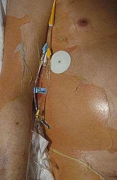
Toxic epidermal necrolysis (TEN), also known as Lyell's syndrome,[1] is a rare, life-threatening skin condition that is usually caused by a reaction to drugs.[2] The disease causes the top layer of skin (the epidermis) to detach from the lower layers of the skin (the dermis), all over the body, leaving the body susceptible to severe infection. The case fatality ratio ranges from 25 to 30%, and death usually occurs as a result of sepsis and subsequent multiorgan system failure. Treatment primarily involves discontinuing the use of causative agent(s), and supportive care in either the intensive care unit or burn unit of a hospital.[3][4]
The incidence of TEN is between 0.4 and 1.9 cases per million each year.[5] TEN exists on a continuum with Stevens–Johnson syndrome (SJS). The condition is called TEN when >30% of the body surface area is involved. It is called SJS when <10% of body surface area is involved. The intermediate form, with 10 to 30 percent body surface area involvement, is called "SJS/TEN."[6][7] Although there was initially debate about whether TEN and SJS fall on a spectrum of disease that includes erythema multiforme (EM), they are now considered separate conditions.[5][8][9]
Signs and symptoms
Prodrome
TEN ultimately results in extensive skin involvement with redness, necrosis, and detachment of the top (epidermal) layer of the skin and mucosa. Before these severe findings develop, people often have a flu-like prodrome, with a cough, runny nose, fever, decreased appetite and malaise. A history of drug exposure exists on average 14 days (ranging from 1–4 weeks) prior to the onset of symptoms, but may result as early as 48 hours if it is a reexposure.[10]
Skin Findings
Initial skin findings include red-purple, dusky, flat spots known as macules that start on the trunk and spread out from there. These skin lesions then transform into large blisters. The affected skin can then become necrotic or sag from the body and peel off in great swaths.[3]
Mucosal Findings
Nearly all people with TEN have oral, eye and genital involvement as well. Painful crusts and erosions may develop on any mucosal surface.[11] The mouth becomes blistered and eroded, making eating difficult and sometimes necessitating feeding through a nasogastric tube through the nose or a gastric tube directly into the stomach. The eyes can become swollen, crusted, and ulcerated, leading to potential blindness. The most common problem with the eyes is severe conjunctivitis.[12]
Cause
Drug reactions have been reported to cause 80-95% of TEN cases.[5]
The drugs most often implicated in TEN are:
- antibiotics
- nonsteroidal anti-inflammatory drugs
- allopurinol
- antimetabolites (methotrexate)
- antiretroviral drugs (nevirapine)
- corticosteroids
- anxiolytics (chlormezanone)
- anticonvulsants (phenobarbital, phenytoin, carbamazepine, and valproic acid).[2]
TEN has also been reported to result from infection with Mycoplasma pneumoniae, dengue virus. Contrast agents used in imaging studies as well as transplantation of bone marrow or organs have also been linked to TEN development.[2][5]
HIV
HIV-positive individuals have 1000 times the risk of developing SJS/TEN compared to the general population. The reason for this increased risk is not clear.[3]
Genetics
Certain genetic factors are associated with increased risk of TEN. For example, certain HLA-types such as, HLA-B*1502,[13] HLA-A*3101,[14] and HLA-B*5801,[15] have been to be linked with TEN development when exposed to specific drugs.
Pathogenesis
The immune system's role in the precise pathogenesis of TEN remains unclear. It appears that a certain type of immune cell (cytotoxic CD8+ T cell) is primarily responsible for keratinocyte death and subsequent skin detachment. Keratinocytes are the cells found lower in the epidermis and specialize in holding the surrounding skin cells together. It is theorized that CD8+ immune cells become overactive by stimulation from drugs or drug metabolites. CD8+ T cells then mediate keratinocyte cell death through release of a number of molecules, including perforin, granzyme B, and granulysin. Other agents, including tumor necrosis factor alpha and fas ligand, also appear to be involved in TEN pathogenesis.[5]
Diagnosis
The diagnosis of TEN is based on both clinical and histologic findings. Early TEN can resemble non-specific drug reactions, so clinicians should maintain a high index of suspicion for TEN. The presence of oral, ocular, and/or genital mucositis is helpful diagnostically, as these findings are present in nearly all patients with TEN. The Nikolsky sign - a separation of the papillary dermis from the basal layer upon gentle lateral pressure - and the Asboe-Hansen sign - a lateral extension of bullae with pressure - are also helpful diagnostic signs found in patients with TEN.[3]
Given the significant morbidity and mortality from TEN, as well as improvement in outcome from prompt treatment, there is significant interest in the discovery of serum biomarkers for early diagnosis of TEN. Serum granulysin and serum high-mobility group protein B1 (HMGB1) are among a few of the markers being investigated which have shown promise in early research.[3]
Histology
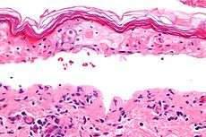
Definitive diagnosis of TEN often requires biopsy confirmation. Histologically, early TEN shows scattered necrotic keratinocytes. In more advanced TEN, full thickness epidermal necrosis is visualized, with a subepidermal split, and scant inflammatory infiltrate in the papillary dermis. Epidermal necrosis found on histology is sensitive but not specific finding for TEN.[3]
-

Confluent Epidermal Necrosis, low mag
-

Confluent Epidermal Necrosis, high mag
Differential Diagnosis
- Staphylococcal scalded skin syndrome
- Drug-induced linear immunoglobulin A dermatosis
- Acute graft versus host disease
- Acute generalized exanthematous pustulosis
- Erythroderma
- Drug reaction with eosinophilia and systemic symptoms aka DRESS
- A generalized morbilliform eruption[16]
Treatment
The primary treatment of TEN is discontinuation of the causative factor(s), usually an offending drug, early referral and management in burn units or intensive care units, supportive management, and nutritional support.[3]
Current literature does not convincingly support use of any adjuvant systemic therapy. Initial interest in Intravenous immunoglobulin (IVIG) came from research showing that IVIG could inhibit Fas-FasL mediated keratinocyte apoptosis in vitro.[17] Unfortunately, research studies reveal conflicting support for use of IVIG in treatment of TEN.[18] Ability to draw more generalized conclusions from research to date has been limited by lack of controlled trials, and inconsistency in study design in terms of disease severity, IVIG dose, and timing of IVIG administration.[3] Larger, high quality trials are needed to assess the actual benefit of IVIG in TEN.
Numerous other adjuvant therapies have been tried in TEN including, corticosteroids, cyclosporin, cyclophosphamide, plasmapheresis, pentoxifylline, N-acetylcysteine, ulinastatin, infliximab, and Granulocyte colony-stimulating factors (if TEN associated-leukopenia exists). There is mixed evidence for use of corticosteriods and scant evidence for the other therapies.[3]
Prognosis
The mortality for toxic epidermal necrolysis is 25-30%.[3] Loss of the skin leaves patients vulnerable to infections from fungi and bacteria, and can result in sepsis, the leading cause of death in the disease.[2] Death is caused either by infection or by respiratory distress which is either due to pneumonia or damage to the linings of the airway. Microscopic analysis of tissue (especially the degree of dermal mononuclear inflammation and the degree of inflammation in general) can play a role in determining the prognosis of individual cases.[19]
Severity score
The "Severity of Illness Score for Toxic Epidermal Necrolysis" (SCORTEN) is a scoring system developed to assess the severity of TEN and predict mortality in patients with acute TEN.[20]
One point is given for each of the following factors
- age>40
- heart rate >120 beats/minute
- carrying diagnosis of cancer
- separation of epidermis on more than ten percent of body surface area (BSA) on day 1.
- Blood Urea Nitrogen >28 mg/dL
- Glucose >252 mg/dL
- Bicarbonate <20mEq/L
Score
- 0-1 - 3.2% mortality
- 2 - 12.2% mortality
- 3 - 35.3% mortality
- 4 - 58.3% mortality
- ≥5 - 90%mortality
Of note, this scoring system is most valuable when used on the first and 3rd day of hospitalization, and it may underestimate mortality in patients with respiratory symptoms.[21]
Long-term complications
Those who survive the acute phase of TEN often suffer long-term complications affecting the skin and eyes. Skin manifestations can include scarring, eruptive melanocytic nevi, vulvovaginal stenosis, and dyspareunia. Ocular symptoms are the most common complication in TEN, experienced by 20-79% of those with TEN, even by those who do not experience immediate ocular manifestations. These can include dry eyes, photophobia, symblepharon, corneal scarring or xerosis, subconjunctival fibrosis, trichiasis, decreased visual acuity, and blindness.[21]
References
- ↑ Rapini, Ronald P.; Bolognia, Jean L.; Jorizzo, Joseph L. (2007). Dermatology: 2-Volume Set. St. Louis: Mosby. ISBN 1-4160-2999-0.
- 1 2 3 4 Garra, GP (2007). "Toxic Epidermal Necrolysis". Emedicine.com. Retrieved on December 13, 2007.
- 1 2 3 4 5 6 7 8 9 10 Schwartz, RA; McDonough, PH; Lee, BW (August 2013). "Toxic epidermal necrolysis: Part II. Prognosis, sequelae, diagnosis, differential diagnosis, prevention, and treatment.". Journal of the American Academy of Dermatology 69 (2): 187.e1–16; quiz 203–4. doi:10.1016/j.jaad.2013.05.002. PMID 23866879.
- ↑ http://www.genome.gov/27560487
- 1 2 3 4 5 Schwartz, RA; McDonough, PH; Lee, BW (August 2013). "Toxic epidermal necrolysis: Part I. Introduction, history, classification, clinical features, systemic manifestations, etiology, and immunopathogenesis.". Journal of the American Academy of Dermatology 69 (2): 173.e1–13; quiz 185–6. doi:10.1016/j.jaad.2013.05.003. PMID 23866878.
- ↑ Nirken, Milton. "Stevens-Johnson syndrome and toxic epidermal necrolysis: Pathogenesis, clinical manifestations, and diagnosis". UpToDate. Wolters Kluwer. Retrieved 21 November 2014.
- ↑ Bastuji-Garin, S; Rzany, B; Stern, RS; Shear, NH; Naldi, L; Roujeau, JC (January 1993). "Clinical classification of cases of toxic epidermal necrolysis, Stevens-Johnson syndrome, and erythema multiforme.". Archives of dermatology 129 (1): 92–6. doi:10.1001/archderm.129.1.92. PMID 8420497.
- ↑ Carrozzo M, Togliatto M, Gandolfo S (1999). "Erythema multiforme. A heterogeneous pathologic phenotype". Minerva Stomatol 48 (5): 217–26. PMID 10434539.
- ↑ Farthing P, Bagan J, Scully C (2005). "Mucosal disease series. Number IV. Erythema multiforme". Oral Dis 11 (5): 261–7. doi:10.1111/j.1601-0825.2005.01141.x. PMID 16120111.
- ↑ Jordan, MH; Lewis, MS; Jeng, JG; Rees, JM (1991). "Treatment of toxic epidermal necrolysis by burn units: another market or another threat?". The Journal of burn care & rehabilitation 12 (6): 579–81. doi:10.1097/00004630-199111000-00015. PMID 1779014.
- ↑ Roujeau, JC; Stern, RS (10 November 1994). "Severe adverse cutaneous reactions to drugs.". The New England Journal of Medicine 331 (19): 1272–85. doi:10.1056/nejm199411103311906. PMID 7794310.
- ↑ Morales, ME; Purdue, GF; Verity, SM; Arnoldo, BD; Blomquist, PH (October 2010). "Ophthalmic Manifestations of Stevens-Johnson Syndrome and Toxic Epidermal Necrolysis and Relation to SCORTEN.". American journal of ophthalmology 150 (4): 505–510.e1. doi:10.1016/j.ajo.2010.04.026. PMID 20619392.
- ↑ Hung, SI; Chung, WH; Jee, SH; Chen, WC; Chang, YT; Lee, WR; Hu, SL; Wu, MT; Chen, GS; Wong, TW; Hsiao, PF; Chen, WH; Shih, HY; Fang, WH; Wei, CY; Lou, YH; Huang, YL; Lin, JJ; Chen, YT (April 2006). "Genetic susceptibility to carbamazepine-induced cutaneous adverse drug reactions.". Pharmacogenetics and genomics 16 (4): 297–306. doi:10.1097/01.fpc.0000199500.46842.4a. PMID 16538176.
- ↑ McCormack, M; Alfirevic, A; Bourgeois, S; Farrell, JJ; Kasperavičiūtė, D; Carrington, M; Sills, GJ; Marson, T; Jia, X; de Bakker, PI; Chinthapalli, K; Molokhia, M; Johnson, MR; O'Connor, GD; Chaila, E; Alhusaini, S; Shianna, KV; Radtke, RA; Heinzen, EL; Walley, N; Pandolfo, M; Pichler, W; Park, BK; Depondt, C; Sisodiya, SM; Goldstein, DB; Deloukas, P; Delanty, N; Cavalleri, GL; Pirmohamed, M (24 March 2011). "HLA-A*3101 and carbamazepine-induced hypersensitivity reactions in Europeans.". The New England Journal of Medicine 364 (12): 1134–43. doi:10.1056/nejmoa1013297. PMID 21428769.
- ↑ Tohkin, M; Kaniwa, N; Saito, Y; Sugiyama, E; Kurose, K; Nishikawa, J; Hasegawa, R; Aihara, M; Matsunaga, K; Abe, M; Furuya, H; Takahashi, Y; Ikeda, H; Muramatsu, M; Ueta, M; Sotozono, C; Kinoshita, S; Ikezawa, Z; Japan Pharmacogenomics Data Science, Consortium (February 2013). "A whole-genome association study of major determinants for allopurinol-related Stevens-Johnson syndrome and toxic epidermal necrolysis in Japanese patients.". The pharmacogenomics journal 13 (1): 60–9. doi:10.1038/tpj.2011.41. PMID 21912425.
- ↑ Schwartz, RA; McDonough, PH; Lee, BW (Aug 2013). "Toxic epidermal necrolysis: Part II. Prognosis, sequelae, diagnosis, differential diagnosis, prevention, and treatment.". Journal of the American Academy of Dermatology 69 (2): 187.e1–16; quiz 203–4. doi:10.1016/j.jaad.2013.05.002. PMID 23866879.
- ↑ Zajicek, R; Pintar, D; Broz, L; Suca, H; Königova, R (May 2012). "Toxic epidermal necrolysis and Stevens-Johnson syndrome at the Prague Burn Centre 1998-2008.". Journal of the European Academy of Dermatology and Venereology : JEADV 26 (5): 639–43. doi:10.1111/j.1468-3083.2011.04143.x. PMID 21668825.
- ↑ Rajaratnam R, Mann C, Balasubramaniam P, et al. (December 2010). "Toxic epidermal necrolysis: retrospective analysis of 21 consecutive cases managed at a tertiary centre". Clin. Exp. Dermatol. 35 (8): 853–62. doi:10.1111/j.1365-2230.2010.03826.x. PMID 20456393.
- ↑ Quinn AM; Brown, K; Bonish, BK; Curry, J; Gordon, KB; Sinacore, J; Gamelli, R; Nickoloff, BJ; et al. (2005). "Uncovering histological criteria with prognostic significance in toxic epidermal necrolysis". Arch Dermatol 141 (6): 683–7. doi:10.1001/archderm.141.6.683. PMID 15967913.
- ↑ Schwartz, RA; McDonough, PH; Lee, BW (August 2013). "Toxic epidermal necrolysis: Part II. Prognosis, sequelae, diagnosis, differential diagnosis, prevention, and treatment.". Journal of the American Academy of Dermatology 69 (2): 187.e1–16; quiz 203–4. doi:10.1016/j.jaad.2013.05.002. PMID 23866879.
- 1 2 3 DeMers, G; Meurer, WJ; Shih, R; Rosenbaum, S; Vilke, GM (December 2012). "Tissue plasminogen activator and stroke: review of the literature for the clinician.". The Journal of emergency medicine 43 (6): 1149–54. doi:10.1016/j.jemermed.2012.05.005. PMID 22818644.
External links
- 18-203e. at Merck Manual of Diagnosis and Therapy Home Edition
- Stevens Johnson Syndrome Foundation
- DermNetNZ
- Understanding Stevens Johnson Syndrome and Toxic Epidermal Necrolysis
| ||||||||||||||||||||||||||||||||||||||||||||