Teres major muscle
| Teres major muscle | |
|---|---|
|
Posterior view showing the relations between teres major muscle (in red) and the other muscles connecting the upper extremity to the vertebral column. | |
 Teres major muscle (in red) seen from back. | |
| Details | |
| Origin | Posterior aspect of the inferior angle of the scapula |
| Insertion | Medial lip of the intertubercular sulcus of the humerus |
| Artery | Subscapular and circumflex scapular arteries |
| Nerve | Lower subscapular nerve (segmental levels C5 and C6) |
| Actions | adduct the humerus, Internal rotation (medial rotation) of the humerus, extend the humerus from flexed position, Protracts scapula, Depress shoulder |
| Identifiers | |
| Latin | Musculus teres major |
| Dorlands /Elsevier | m_22/12551115 |
| TA | A04.6.02.011 |
| FMA | 32549 |
The teres major muscle (Latin teres meaning 'rounded') is a muscle of the upper limb and one of seven scapulohumeral muscles. It is a thick but somewhat flattened muscle, innervated by the lower subscapular nerve (C5 and C6).
Structure
It arises from the oval area on the dorsal surface of the inferior angle of the scapula, and from the fibrous septa interposed between this muscle and the rotator cuff lateral rotator pair of the teres minor and infraspinatus.
The fibers of teres major insert into the medial lip of the intertubercular sulcus of the humerus.
Relations
The tendon, at its insertion, lies behind that of the latissimus dorsi, from which it is separated by a bursa, the two tendons being, however, united along their lower borders for a short distance.
Together with teres minor muscle, teres major muscle forms the axillary space, through which several important arteries and veins pass.
The teres major muscle is innervated by the lower subscapular nerve of the brachial plexus.
Function
The teres major is a medial rotator and adductor of the humerus and assists the latissimus dorsi in drawing the previously raised humerus downward and backward (extension, but not hyper extension). It also helps stabilize the humeral head in the glenoid cavity.
Additional images
-

Position of teres major muscle (shown in red). Animation.
-

Muscles on the dorsum of the scapula, and the Triceps brachii muscle:
#3 latissimus dorsi muscle
#5 teres major muscle
#6 teres minor muscle
#7 supraspinatus muscle
#8 infraspinatus muscle
#13 long head of triceps brachii muscle -

Surface anatomy of the back. (Label for Teres major at upper right.)
-

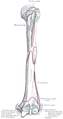
Left humerus. Anterior view.
-
Teres major muscle
-

Left scapula. Posterior surface.
-
Teres major muscle
See also
References
This article incorporates text in the public domain from the 20th edition of Gray's Anatomy (1918)
External links
| Wikimedia Commons has media related to Teres major muscles. |
- 711655502 at GPnotebook
- Anatomy figure: 03:03-06 at Human Anatomy Online, SUNY Downstate Medical Center
- PTCentral
| ||||||||||||||||||||||||||||||||||||||||||||||||||||||||||||||||||||||||||||||||||||