RNA

Ribonucleic acid (RNA) is a polymeric molecule implicated in various biological roles in coding, decoding, regulation, and expression of genes. RNA and DNA are nucleic acids, and, along with proteins and carbohydrates, constitute the three major macromolecules essential for all known forms of life. Like DNA, RNA is assembled as a chain of nucleotides, but unlike DNA it is more often found in nature as a single-strand folded onto itself, rather than a paired double-strand. Cellular organisms use messenger RNA (mRNA) to convey genetic information (using the letters G, U, A, and C to denote the nitrogenous bases guanine, uracil, adenine, and cytosine) that directs synthesis of specific proteins. Many viruses encode their genetic information using an RNA genome.
Some RNA molecules play an active role within cells by catalyzing biological reactions, controlling gene expression, or sensing and communicating responses to cellular signals. One of these active processes is protein synthesis, a universal function wherein mRNA molecules direct the assembly of proteins on ribosomes. This process uses transfer RNA (tRNA) molecules to deliver amino acids to the ribosome, where ribosomal RNA (rRNA) then links amino acids together to form proteins.
Comparison with DNA


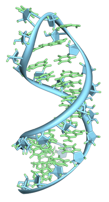
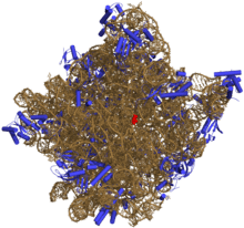
Like DNA, most biologically active RNAs, including mRNA, tRNA, rRNA, snRNAs, and other non-coding RNAs, contain self-complementary sequences that allow parts of the RNA to fold[5] and pair with itself to form double helices. Analysis of these RNAs has revealed that they are highly structured. Unlike DNA, their structures do not consist of long double helices, but rather collections of short helices packed together into structures akin to proteins. In this fashion, RNAs can achieve chemical catalysis, like enzymes.[6] For instance, determination of the structure of the ribosome—an enzyme that catalyzes peptide bond formation—revealed that its active site is composed entirely of RNA.[7]
Structure

Each nucleotide in RNA contains a ribose sugar, with carbons numbered 1' through 5'. A base is attached to the 1' position, in general, adenine (A), cytosine (C), guanine (G), or uracil (U). Adenine and guanine are purines, cytosine and uracil are pyrimidines. A phosphate group is attached to the 3' position of one ribose and the 5' position of the next. The phosphate groups do not have a negative charge each at physiological pH, making RNA a charged molecule (polyanion). The bases form hydrogen bonds between cytosine and guanine, between adenine and uracil and between guanine and uracil.[8] However, other interactions are possible, such as a group of adenine bases binding to each other in a bulge,[9] or the GNRA tetraloop that has a guanine–adenine base-pair.[8]
An important structural feature of RNA that distinguishes it from DNA is the presence of a hydroxyl group at the 2' position of the ribose sugar. The presence of this functional group causes the helix to mostly adopt the A-form geometry,[10] although in single strand dinucleotide contexts, RNA can rarely also adopt the B-form most commonly observed in DNA.[11] The A-form geometry results in a very deep and narrow major groove and a shallow and wide minor groove.[12] A second consequence of the presence of the 2'-hydroxyl group is that in conformationally flexible regions of an RNA molecule (that is, not involved in formation of a double helix), it can chemically attack the adjacent phosphodiester bond to cleave the backbone.[13]
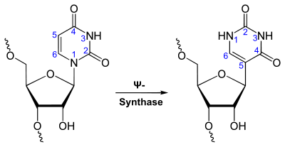
RNA is transcribed with only four bases (adenine, cytosine, guanine and uracil),[14] but these bases and attached sugars can be modified in numerous ways as the RNAs mature. Pseudouridine (Ψ), in which the linkage between uracil and ribose is changed from a C–N bond to a C–C bond, and ribothymidine (T) are found in various places (the most notable ones being in the TΨC loop of tRNA).[15] Another notable modified base is hypoxanthine, a deaminated adenine base whose nucleoside is called inosine (I). Inosine plays a key role in the wobble hypothesis of the genetic code.[16]
There are more than 100 other naturally occurring modified nucleosides,[17] The greatest structural diversity of modifications can be found in tRNA,[18] while pseudouridine and nucleosides with 2'-O-methylribose often present in rRNA are the most common.[19] The specific roles of many of these modifications in RNA are not fully understood. However, it is notable that, in ribosomal RNA, many of the post-transcriptional modifications occur in highly functional regions, such as the peptidyl transferase center and the subunit interface, implying that they are important for normal function.[20]
The functional form of single-stranded RNA molecules, just like proteins, frequently requires a specific tertiary structure. The scaffold for this structure is provided by secondary structural elements that are hydrogen bonds within the molecule. This leads to several recognizable "domains" of secondary structure like hairpin loops, bulges, and internal loops.[21] Since RNA is charged, metal ions such as Mg2+ are needed to stabilise many secondary and tertiary structures.[22]
The naturally occurring enantiomer of RNA is D-RNA composed of D-ribonucleotides. All chirality centers are located in the D-ribose. By the use of L-ribose or rather L-ribonucleotides, L-RNA can be synthesized. L-RNA is much more stable against degradation by RNase.[23]
Like other structured biopolymers such as proteins, one can define topology of a folded RNA molecule. This is often done based on arrangement of intra-chain contacts within a folded RNA, termed as circuit topology.
Synthesis
Synthesis of RNA is usually catalyzed by an enzyme—RNA polymerase—using DNA as a template, a process known as transcription. Initiation of transcription begins with the binding of the enzyme to a promoter sequence in the DNA (usually found "upstream" of a gene). The DNA double helix is unwound by the helicase activity of the enzyme. The enzyme then progresses along the template strand in the 3’ to 5’ direction, synthesizing a complementary RNA molecule with elongation occurring in the 5’ to 3’ direction. The DNA sequence also dictates where termination of RNA synthesis will occur.[24]
Primary transcript RNAs are often modified by enzymes after transcription. For example, a poly(A) tail and a 5' cap are added to eukaryotic pre-mRNA and introns are removed by the spliceosome.
There are also a number of RNA-dependent RNA polymerases that use RNA as their template for synthesis of a new strand of RNA. For instance, a number of RNA viruses (such as poliovirus) use this type of enzyme to replicate their genetic material.[25] Also, RNA-dependent RNA polymerase is part of the RNA interference pathway in many organisms.[26]
Types of RNA
Overview

Messenger RNA (mRNA) is the RNA that carries information from DNA to the ribosome, the sites of protein synthesis (translation) in the cell. The coding sequence of the mRNA determines the amino acid sequence in the protein that is produced.[27] However, many RNAs do not code for protein (about 97% of the transcriptional output is non-protein-coding in eukaryotes[28][29][30][31]).
These so-called non-coding RNAs ("ncRNA") can be encoded by their own genes (RNA genes), but can also derive from mRNA introns.[32] The most prominent examples of non-coding RNAs are transfer RNA (tRNA) and ribosomal RNA (rRNA), both of which are involved in the process of translation.[4] There are also non-coding RNAs involved in gene regulation, RNA processing and other roles. Certain RNAs are able to catalyse chemical reactions such as cutting and ligating other RNA molecules,[33] and the catalysis of peptide bond formation in the ribosome;[7] these are known as ribozymes.
In translation
Messenger RNA (mRNA) carries information about a protein sequence to the ribosomes, the protein synthesis factories in the cell. It is coded so that every three nucleotides (a codon) correspond to one amino acid. In eukaryotic cells, once precursor mRNA (pre-mRNA) has been transcribed from DNA, it is processed to mature mRNA. This removes its introns—non-coding sections of the pre-mRNA. The mRNA is then exported from the nucleus to the cytoplasm, where it is bound to ribosomes and translated into its corresponding protein form with the help of tRNA. In prokaryotic cells, which do not have nucleus and cytoplasm compartments, mRNA can bind to ribosomes while it is being transcribed from DNA. After a certain amount of time the message degrades into its component nucleotides with the assistance of ribonucleases.[27]
Transfer RNA (tRNA) is a small RNA chain of about 80 nucleotides that transfers a specific amino acid to a growing polypeptide chain at the ribosomal site of protein synthesis during translation. It has sites for amino acid attachment and an anticodon region for codon recognition that binds to a specific sequence on the messenger RNA chain through hydrogen bonding.[32]
Ribosomal RNA (rRNA) is the catalytic component of the ribosomes. Eukaryotic ribosomes contain four different rRNA molecules: 18S, 5.8S, 28S and 5S rRNA. Three of the rRNA molecules are synthesized in the nucleolus, and one is synthesized elsewhere. In the cytoplasm, ribosomal RNA and protein combine to form a nucleoprotein called a ribosome. The ribosome binds mRNA and carries out protein synthesis. Several ribosomes may be attached to a single mRNA at any time.[27] Nearly all the RNA found in a typical eukaryotic cell is rRNA.
Transfer-messenger RNA (tmRNA) is found in many bacteria and plastids. It tags proteins encoded by mRNAs that lack stop codons for degradation and prevents the ribosome from stalling.[34]
Regulatory RNAs
Several types of RNA can downregulate gene expression by being complementary to a part of an mRNA or a gene's DNA.[35][36] MicroRNAs (miRNA; 21-22 nt) are found in eukaryotes and act through RNA interference (RNAi), where an effector complex of miRNA and enzymes can cleave complementary mRNA, block the mRNA from being translated, or accelerate its degradation.[37][38]
While small interfering RNAs (siRNA; 20-25 nt) are often produced by breakdown of viral RNA, there are also endogenous sources of siRNAs.[39][40] siRNAs act through RNA interference in a fashion similar to miRNAs. Some miRNAs and siRNAs can cause genes they target to be methylated, thereby decreasing or increasing transcription of those genes.[41][42][43] Animals have Piwi-interacting RNAs (piRNA; 29-30 nt) that are active in germline cells and are thought to be a defense against transposons and play a role in gametogenesis.[44][45]
Many prokaryotes have CRISPR RNAs, a regulatory system similar to RNA interference.[46] Antisense RNAs are widespread; most downregulate a gene, but a few are activators of transcription.[47] One way antisense RNA can act is by binding to an mRNA, forming double-stranded RNA that is enzymatically degraded.[48] There are many long noncoding RNAs that regulate genes in eukaryotes,[49] one such RNA is Xist, which coats one X chromosome in female mammals and inactivates it.[50]
An mRNA may contain regulatory elements itself, such as riboswitches, in the 5' untranslated region or 3' untranslated region; these cis-regulatory elements regulate the activity of that mRNA.[51] The untranslated regions can also contain elements that regulate other genes.[52]
In RNA processing

Many RNAs are involved in modifying other RNAs. Introns are spliced out of pre-mRNA by spliceosomes, which contain several small nuclear RNAs (snRNA),[4] or the introns can be ribozymes that are spliced by themselves.[53] RNA can also be altered by having its nucleotides modified to other nucleotides than A, C, G and U. In eukaryotes, modifications of RNA nucleotides are in general directed by small nucleolar RNAs (snoRNA; 60-300 nt),[32] found in the nucleolus and cajal bodies. snoRNAs associate with enzymes and guide them to a spot on an RNA by basepairing to that RNA. These enzymes then perform the nucleotide modification. rRNAs and tRNAs are extensively modified, but snRNAs and mRNAs can also be the target of base modification.[54][55] RNA can also be methylated.[56][57]
RNA genomes
Like DNA, RNA can carry genetic information. RNA viruses have genomes composed of RNA that encodes a number of proteins. The viral genome is replicated by some of those proteins, while other proteins protect the genome as the virus particle moves to a new host cell. Viroids are another group of pathogens, but they consist only of RNA, do not encode any protein and are replicated by a host plant cell's polymerase.[58]
In reverse transcription
Reverse transcribing viruses replicate their genomes by reverse transcribing DNA copies from their RNA; these DNA copies are then transcribed to new RNA. Retrotransposons also spread by copying DNA and RNA from one another,[59] and telomerase contains an RNA that is used as template for building the ends of eukaryotic chromosomes.[60]
Double-stranded RNA
Double-stranded RNA (dsRNA) is RNA with two complementary strands, similar to the DNA found in all cells. dsRNA forms the genetic material of some viruses (double-stranded RNA viruses). Double-stranded RNA such as viral RNA or siRNA can trigger RNA interference in eukaryotes, as well as interferon response in vertebrates.[61][62][63][64]
Key discoveries in RNA biology

Research on RNA has led to many important biological discoveries and numerous Nobel Prizes. Nucleic acids were discovered in 1868 by Friedrich Miescher, who called the material 'nuclein' since it was found in the nucleus.[65] It was later discovered that prokaryotic cells, which do not have a nucleus, also contain nucleic acids. The role of RNA in protein synthesis was suspected already in 1939.[66] Severo Ochoa won the 1959 Nobel Prize in Medicine (shared with Arthur Kornberg) after he discovered an enzyme that can synthesize RNA in the laboratory.[67] However, the enzyme discovered by Ochoa (polynucleotide phosphorylase) was later shown to be responsible for RNA degradation, not RNA synthesis. In 1956 Alex Rich and David Davies hybridized two separate strands of RNA to form the first crystal of RNA whose structure could be determined by X-ray crystallography.[68]
The sequence of the 77 nucleotides of a yeast tRNA was found by Robert W. Holley in 1965,[69] winning Holley the 1968 Nobel Prize in Medicine (shared with Har Gobind Khorana and Marshall Nirenberg). In 1967, Carl Woese hypothesized that RNA might be catalytic and suggested that the earliest forms of life (self-replicating molecules) could have relied on RNA both to carry genetic information and to catalyze biochemical reactions—an RNA world.[70][71]
During the early 1970s, retroviruses and reverse transcriptase were discovered, showing for the first time that enzymes could copy RNA into DNA (the opposite of the usual route for transmission of genetic information). For this work, David Baltimore, Renato Dulbecco and Howard Temin were awarded a Nobel Prize in 1975. In 1976, Walter Fiers and his team determined the first complete nucleotide sequence of an RNA virus genome, that of bacteriophage MS2.[72]
In 1977, introns and RNA splicing were discovered in both mammalian viruses and in cellular genes, resulting in a 1993 Nobel to Philip Sharp and Richard Roberts. Catalytic RNA molecules (ribozymes) were discovered in the early 1980s, leading to a 1989 Nobel award to Thomas Cech and Sidney Altman. In 1990, it was found in Petunia that introduced genes can silence similar genes of the plant's own, now known to be a result of RNA interference.[73][74]
At about the same time, 22 nt long RNAs, now called microRNAs, were found to have a role in the development of C. elegans.[75] Studies on RNA interference gleaned a Nobel Prize for Andrew Fire and Craig Mello in 2006, and another Nobel was awarded for studies on the transcription of RNA to Roger Kornberg in the same year. The discovery of gene regulatory RNAs has led to attempts to develop drugs made of RNA, such as siRNA, to silence genes.[76]
Evolution
In March 2015, complex DNA and RNA organic compounds of life, including uracil, cytosine and thymine, were reportedly formed in the laboratory under outer space conditions, using starting chemicals, such as pyrimidine, found in meteorites. Pyrimidine, like polycyclic aromatic hydrocarbons (PAHs), the most carbon-rich chemical found in the Universe, may have been formed in red giants or in interstellar dust and gas clouds, according to the scientists.[77]
See also
- Biomolecular structure
- DNA, RNA and proteins: The three essential macromolecules of life
- DNA
- History of RNA Biology
- List of RNA Biologists
References
- ↑ "RNA: The Versatile Molecule". University of Utah. 2015.
- ↑ "Nucleotides and Nucleic Acids" (PDF). University of California, Los Angeles.
- ↑ R.N. Shukla. Analysis of Chromosomes. ISBN 9789384568177.
- 1 2 3 Berg JM; Tymoczko JL; Stryer L (2002). Biochemistry (5th ed.). WH Freeman and Company. pp. 118–19, 781–808. ISBN 0-7167-4684-0. OCLC 179705944 48055706 59502128.
- ↑ I. Tinoco & C. Bustamante (1999). "How RNA folds". J. Mol. Biol. 293 (2): 271–281. doi:10.1006/jmbi.1999.3001. PMID 10550208.
- ↑ Higgs PG (2000). "RNA secondary structure: physical and computational aspects". Quarterly Reviews of Biophysics 33 (3): 199–253. doi:10.1017/S0033583500003620. PMID 11191843.
- 1 2 Nissen P; Hansen J; Ban N; Moore PB; Steitz TA (2000). "The structural basis of ribosome activity in peptide bond synthesis". Science 289 (5481): 920–30. Bibcode:2000Sci...289..920N. doi:10.1126/science.289.5481.920. PMID 10937990.
- 1 2 Lee JC; Gutell RR (2004). "Diversity of base-pair conformations and their occurrence in rRNA structure and RNA structural motifs". J. Mol. Biol. 344 (5): 1225–49. doi:10.1016/j.jmb.2004.09.072. PMID 15561141.
- ↑ Barciszewski J; Frederic B; Clark C (1999). RNA biochemistry and biotechnology. Springer. pp. 73–87. ISBN 0-7923-5862-7. OCLC 52403776.
- ↑ Salazar M; Fedoroff OY; Miller JM; Ribeiro NS; Reid BR (1992). "The DNA strand in DNAoRNA hybrid duplexes is neither B-form nor A-form in solution". Biochemistry 32 (16): 4207–15. doi:10.1021/bi00067a007. PMID 7682844.
- ↑ Sedova A; Banavali NK (2016). "RNA approaches the B-form in stacked single strand dinucleotide contexts". Biopolymers 105 (2): 65–82. doi:10.1002/bip.22750. PMID 26443416.
- ↑ Hermann T; Patel DJ (2000). "RNA bulges as architectural and recognition motifs". Structure 8 (3): R47–R54. doi:10.1016/S0969-2126(00)00110-6. PMID 10745015.
- ↑ Mikkola S; Stenman E; Nurmi K; Yousefi-Salakdeh E; Strömberg R; Lönnberg H (1999). "The mechanism of the metal ion promoted cleavage of RNA phosphodiester bonds involves a general acid catalysis by the metal aquo ion on the departure of the leaving group". Perkin transactions 2 (8): 1619–26. doi:10.1039/a903691a.
- ↑ Jankowski JAZ; Polak JM (1996). Clinical gene analysis and manipulation: Tools, techniques and troubleshooting. Cambridge University Press. p. 14. ISBN 0-521-47896-0. OCLC 33838261.
- ↑ Yu Q; Morrow CD (2001). "Identification of critical elements in the tRNA acceptor stem and TΨC loop necessary for human immunodeficiency virus type 1 infectivity". J Virol 75 (10): 4902–6. doi:10.1128/JVI.75.10.4902-4906.2001. PMC 114245. PMID 11312362.
- ↑ Elliott MS; Trewyn RW (1983). "Inosine biosynthesis in transfer RNA by an enzymatic insertion of hypoxanthine". J. Biol. Chem. 259 (4): 2407–10. PMID 6365911.
- ↑ Cantara, WA; Crain, PF; Rozenski, J; McCloskey, JA; Harris, KA; Zhang, X; Vendeix, FA; Fabris, D; Agris, PF (January 2011). "The RNA Modification Database, RNAMDB: 2011 update". Nucleic Acids Research 39 (Database issue): D195–201. doi:10.1093/nar/gkq1028. PMC 3013656. PMID 21071406.
- ↑ Söll D; RajBhandary U (1995). TRNA: Structure, biosynthesis, and function. ASM Press. p. 165. ISBN 1-55581-073-X. OCLC 183036381 30663724.
- ↑ Kiss T (2001). "Small nucleolar RNA-guided post-transcriptional modification of cellular RNAs". The EMBO Journal 20 (14): 3617–22. doi:10.1093/emboj/20.14.3617. PMC 125535. PMID 11447102.
- ↑ King TH; Liu B; McCully RR; Fournier MJ (2002). "Ribosome structure and activity are altered in cells lacking snoRNPs that form pseudouridines in the peptidyl transferase center". Molecular Cell 11 (2): 425–35. doi:10.1016/S1097-2765(03)00040-6. PMID 12620230.
- ↑ Mathews DH; Disney MD; Childs JL; Schroeder SJ; Zuker M; Turner DH (2004). "Incorporating chemical modification constraints into a dynamic programming algorithm for prediction of RNA secondary structure". Proc. Natl. Acad. Sci. USA 101 (19): 7287–92. Bibcode:2004PNAS..101.7287M. doi:10.1073/pnas.0401799101. PMC 409911. PMID 15123812.
- ↑ Tan ZJ; Chen SJ (2008). "Salt dependence of nucleic acid hairpin stability". Biophys. J. 95 (2): 738–52. Bibcode:2008BpJ....95..738T. doi:10.1529/biophysj.108.131524. PMC 2440479. PMID 18424500.
- ↑ Vater A; Klussmann S (January 2015). "Turning mirror-image oligonucleotides into drugs: the evolution of Spiegelmer therapeutics". Drug Discovery Today 20 (1): 147–155. doi:10.1016/j.drudis.2014.09.004. PMID 25236655.
- ↑ Nudler E; Gottesman ME (2002). "Transcription termination and anti-termination in E. coli". Genes to Cells 7 (8): 755–68. doi:10.1046/j.1365-2443.2002.00563.x. PMID 12167155.
- ↑ Jeffrey L Hansen; Alexander M Long; Steve C Schultz (1997). "Structure of the RNA-dependent RNA polymerase of poliovirus". Structure 5 (8): 1109–22. doi:10.1016/S0969-2126(97)00261-X. PMID 9309225.
- ↑ Ahlquist P (2002). "RNA-Dependent RNA Polymerases, Viruses, and RNA Silencing". Science 296 (5571): 1270–73. Bibcode:2002Sci...296.1270A. doi:10.1126/science.1069132. PMID 12016304.
- 1 2 3 Cooper GC; Hausman RE (2004). The Cell: A Molecular Approach (3rd ed.). Sinauer. pp. 261–76, 297, 339–44. ISBN 0-87893-214-3. OCLC 174924833 52121379 52359301 56050609.
- ↑ Mattick JS; Gagen MJ (1 September 2001). "The evolution of controlled multitasked gene networks: the role of introns and other noncoding RNAs in the development of complex organisms". Mol. Biol. Evol. 18 (9): 1611–30. doi:10.1093/oxfordjournals.molbev.a003951. PMID 11504843.
- ↑ Mattick, JS (2001). "Noncoding RNAs: the architects of eukaryotic complexity". EMBO Reports 2 (11): 986–91. doi:10.1093/embo-reports/kve230. PMC 1084129. PMID 11713189.
- ↑ Mattick JS (October 2003). "Challenging the dogma: the hidden layer of non-protein-coding RNAs in complex organisms" (PDF). BioEssays : News and Reviews in Molecular, Cellular and Developmental Biology 25 (10): 930–9. doi:10.1002/bies.10332. PMID 14505360.
- ↑ Mattick JS (October 2004). "The hidden genetic program of complex organisms". Scientific American 291 (4): 60–7. doi:10.1038/scientificamerican1004-60. PMID 15487671. Archived from the original on February 8, 2015.
- 1 2 3 Wirta W (2006). Mining the transcriptome – methods and applications. Stockholm: School of Biotechnology, Royal Institute of Technology. ISBN 91-7178-436-5. OCLC 185406288.
- ↑ Rossi JJ (2004). "Ribozyme diagnostics comes of age". Chemistry & Biology 11 (7): 894–95. doi:10.1016/j.chembiol.2004.07.002. PMID 15271347.
- ↑ Gueneau de Novoa P, Williams KP; Williams (2004). "The tmRNA website: reductive evolution of tmRNA in plastids and other endosymbionts". Nucleic Acids Res 32 (Database issue): D104–8. doi:10.1093/nar/gkh102. PMC 308836. PMID 14681369.
- ↑ Carthew, RW; Sontheimer, EJ. "Origins and Mechanisms of miRNAs and siRNAs.". Cell. doi:10.1016/j.cell.2009.01.035. PMC 2675692. PMID 19239886. Retrieved 2009-02-20.
- ↑ Liang, Kung-Hao; Yeh, Chau-Ting. "A gene expression restriction network mediated by sense and antisense Alu sequences located on protein-coding messenger RNAs.". BMC Genomics. doi:10.1186/1471-2164-14-325. PMC 3655826. PMID 23663499. Retrieved 2013-05-11.
- ↑ Wu L; Belasco JG (January 2008). "Let me count the ways: mechanisms of gene regulation by miRNAs and siRNAs". Mol. Cell 29 (1): 1–7. doi:10.1016/j.molcel.2007.12.010. PMID 18206964.
- ↑ Matzke MA; Matzke AJM (2004). "Planting the seeds of a new paradigm". PLoS Biology 2 (5): e133. doi:10.1371/journal.pbio.0020133. PMC 406394. PMID 15138502.
- ↑ Vazquez F; Vaucheret H; Rajagopalan R; Lepers C; Gasciolli V; Mallory AC; Hilbert J; Bartel DP; Crété P (2004). "Endogenous trans-acting siRNAs regulate the accumulation of Arabidopsis mRNAs". Molecular Cell 16 (1): 69–79. doi:10.1016/j.molcel.2004.09.028. PMID 15469823.
- ↑ Watanabe T, Totoki Y, Toyoda A, et al. (May 2008). "Endogenous siRNAs from naturally formed dsRNAs regulate transcripts in mouse oocytes". Nature 453 (7194): 539–43. Bibcode:2008Natur.453..539W. doi:10.1038/nature06908. PMID 18404146.
- ↑ Sontheimer EJ; Carthew RW (July 2005). "Silence from within: endogenous siRNAs and miRNAs". Cell 122 (1): 9–12. doi:10.1016/j.cell.2005.06.030. PMID 16009127.
- ↑ Doran G (2007). "RNAi – Is one suffix sufficient?". Journal of RNAi and Gene Silencing 3 (1): 217–19.
- ↑ Pushparaj PN; Aarthi JJ; Kumar SD; Manikandan J (2008). "RNAi and RNAa — The Yin and Yang of RNAome". Bioinformation 2 (6): 235–7. doi:10.6026/97320630002235. PMC 2258431. PMID 18317570.
- ↑ Horwich MD; Li C; Matranga C; Vagin V; Farley G; Wang P; Zamore PD (2007). "The Drosophila RNA methyltransferase, DmHen1, modifies germline piRNAs and single-stranded siRNAs in RISC". Current Biology 17 (14): 1265–72. doi:10.1016/j.cub.2007.06.030. PMID 17604629.
- ↑ Girard A; Sachidanandam R; Hannon GJ; Carmell MA (2006). "A germline-specific class of small RNAs binds mammalian Piwi proteins". Nature 442 (7099): 199–202. Bibcode:2006Natur.442..199G. doi:10.1038/nature04917. PMID 16751776.
- ↑ Horvath P; Barrangou R (2010). "CRISPR/Cas, the Immune System of Bacteria and Archaea". Science 327 (5962): 167–70. Bibcode:2010Sci...327..167H. doi:10.1126/science.1179555. PMID 20056882.
- ↑ Wagner EG; Altuvia S; Romby P (2002). "Antisense RNAs in bacteria and their genetic elements". Adv Genet. Advances in Genetics 46: 361–98. doi:10.1016/S0065-2660(02)46013-0. ISBN 9780120176465. PMID 11931231.
- ↑ Gilbert SF (2003). Developmental Biology (7th ed.). Sinauer. pp. 101–3. ISBN 0-87893-258-5. OCLC 154656422 154663147 174530692 177000492 177316159 51544170 54743254 59197768 61404850 66754122.
- ↑ Amaral PP; Mattick JS (October 2008). "Noncoding RNA in development". Mammalian genome : official journal of the International Mammalian Genome Society 19 (7–8): 454–92. doi:10.1007/s00335-008-9136-7. PMID 18839252.
- ↑ Heard E; Mongelard F; Arnaud D; Chureau C; Vourc'h C; Avner P (1999). "Human XIST yeast artificial chromosome transgenes show partial X inactivation center function in mouse embryonic stem cells". Proc. Natl. Acad. Sci. USA 96 (12): 6841–46. Bibcode:1999PNAS...96.6841H. doi:10.1073/pnas.96.12.6841. PMC 22003. PMID 10359800.
- ↑ Batey RT (2006). "Structures of regulatory elements in mRNAs". Curr. Opin. Struct. Biol. 16 (3): 299–306. doi:10.1016/j.sbi.2006.05.001. PMID 16707260.
- ↑ Scotto L; Assoian RK (June 1993). "A GC-rich domain with bifunctional effects on mRNA and protein levels: implications for control of transforming growth factor beta 1 expression". Mol. Cell. Biol. 13 (6): 3588–97. PMC 359828. PMID 8497272.
- ↑ Steitz TA; Steitz JA (1993). "A general two-metal-ion mechanism for catalytic RNA". Proc. Natl. Acad. Sci. U.S.A. 90 (14): 6498–502. Bibcode:1993PNAS...90.6498S. doi:10.1073/pnas.90.14.6498. PMC 46959. PMID 8341661.
- ↑ Xie J; Zhang M; Zhou T; Hua X; Tang L; Wu W (2007). "Sno/scaRNAbase: a curated database for small nucleolar RNAs and cajal body-specific RNAs". Nucleic Acids Res 35 (Database issue): D183–7. doi:10.1093/nar/gkl873. PMC 1669756. PMID 17099227.
- ↑ Omer AD; Ziesche S; Decatur WA; Fournier MJ; Dennis PP (2003). "RNA-modifying machines in archaea". Molecular Microbiology 48 (3): 617–29. doi:10.1046/j.1365-2958.2003.03483.x. PMID 12694609.
- ↑ Cavaillé J; Nicoloso M; Bachellerie JP (1996). "Targeted ribose methylation of RNA in vivo directed by tailored antisense RNA guides". Nature 383 (6602): 732–5. Bibcode:1996Natur.383..732C. doi:10.1038/383732a0. PMID 8878486.
- ↑ Kiss-László Z; Henry Y; Bachellerie JP; Caizergues-Ferrer M; Kiss T (1996). "Site-specific ribose methylation of preribosomal RNA: a novel function for small nucleolar RNAs". Cell 85 (7): 1077–88. doi:10.1016/S0092-8674(00)81308-2. PMID 8674114.
- ↑ Daròs JA; Elena SF; Flores R (2006). "Viroids: an Ariadne's thread into the RNA labyrinth". EMBO Rep 7 (6): 593–8. doi:10.1038/sj.embor.7400706. PMC 1479586. PMID 16741503.
- ↑ Kalendar R; Vicient CM; Peleg O; Anamthawat-Jonsson K; Bolshoy A; Schulman AH (2004). "Large retrotransposon derivatives: abundant, conserved but nonautonomous retroelements of barley and related genomes". Genetics 166 (3): 1437–50. doi:10.1534/genetics.166.3.1437. PMC 1470764. PMID 15082561.
- ↑ Podlevsky JD; Bley CJ; Omana RV; Qi X; Chen JJ (2008). "The telomerase database". Nucleic Acids Res 36 (Database issue): D339–43. doi:10.1093/nar/gkm700. PMC 2238860. PMID 18073191.
- ↑ Blevins, T.; Rajeswaran, R.; Shivaprasad, PV.; Beknazariants, D.; Si-Ammour, A.; Park, HS.; Vazquez, F.; Robertson, D.; Meins, F. (2006). "Four plant Dicers mediate viral small RNA biogenesis and DNA virus induced silencing". Nucleic Acids Res 34 (21): 6233–46. doi:10.1093/nar/gkl886. PMC 1669714. PMID 17090584.
- ↑ Jana S; Chakraborty C; Nandi S; Deb JK (2004). "RNA interference: potential therapeutic targets". Appl. Microbiol. Biotechnol. 65 (6): 649–57. doi:10.1007/s00253-004-1732-1. PMID 15372214.
- ↑ Schultz U; Kaspers B; Staeheli P (2004). "The interferon system of non-mammalian vertebrates". Dev. Comp. Immunol. 28 (5): 499–508. doi:10.1016/j.dci.2003.09.009. PMID 15062646.
- ↑ Whitehead, K. A.; Dahlman, J. E.; Langer, R. S.; Anderson, D. G. (2011). "Silencing or Stimulation? SiRNA Delivery and the Immune System". Annual Review of Chemical and Biomolecular Engineering 2: 77–96. doi:10.1146/annurev-chembioeng-061010-114133. PMID 22432611.
- ↑ Dahm R (2005). "Friedrich Miescher and the discovery of DNA". Developmental Biology 278 (2): 274–88. doi:10.1016/j.ydbio.2004.11.028. PMID 15680349.
- ↑ Caspersson T; Schultz J (1939). "Pentose nucleotides in the cytoplasm of growing tissues". Nature 143 (3623): 602–3. Bibcode:1939Natur.143..602C. doi:10.1038/143602c0.
- ↑ Ochoa S (1959). "Enzymatic synthesis of ribonucleic acid" (PDF). Nobel Lecture.
- ↑ Rich A; Davies, D (1956). "A New Two-Stranded Helical Structure: Polyadenylic Acid and Polyuridylic Acid". Journal of the American Chemical Society 78 (14): 3548–3549. doi:10.1021/ja01595a086.
- ↑ Holley RW, et al. (1965). "Structure of a ribonucleic acid". Science 147 (3664): 1462–65. Bibcode:1965Sci...147.1462H. doi:10.1126/science.147.3664.1462. PMID 14263761.
- ↑ Siebert S (2006). "Common sequence structure properties and stable regions in RNA secondary structures" (PDF). Dissertation, Albert-Ludwigs-Universität, Freiburg im Breisgau. p. 1. Archived from the original (PDF) on March 9, 2012.
- ↑ Szathmáry E (1999). "The origin of the genetic code: amino acids as cofactors in an RNA world". Trends Genet 15 (6): 223–9. doi:10.1016/S0168-9525(99)01730-8. PMID 10354582.
- ↑ Fiers W, et al. (1976). "Complete nucleotide-sequence of bacteriophage MS2-RNA: primary and secondary structure of replicase gene". Nature 260 (5551): 500–7. Bibcode:1976Natur.260..500F. doi:10.1038/260500a0. PMID 1264203.
- ↑ Napoli C; Lemieux C; Jorgensen R (1990). "Introduction of a chimeric chalcone synthase gene into petunia results in reversible co-suppression of homologous genes in trans". Plant Cell 2 (4): 279–89. doi:10.1105/tpc.2.4.279. PMC 159885. PMID 12354959.
- ↑ Dafny-Yelin M; Chung SM; Frankman EL; Tzfira T (December 2007). "pSAT RNA interference vectors: a modular series for multiple gene down-regulation in plants". Plant Physiol. 145 (4): 1272–81. doi:10.1104/pp.107.106062. PMC 2151715. PMID 17766396.
- ↑ Ruvkun G (2001). "Glimpses of a tiny RNA world". Science 294 (5543): 797–99. doi:10.1126/science.1066315. PMID 11679654.
- ↑ Fichou Y; Férec C (2006). "The potential of oligonucleotides for therapeutic applications". Trends in Biotechnology 24 (12): 563–70. doi:10.1016/j.tibtech.2006.10.003. PMID 17045686.
- ↑ Marlaire, Ruth (3 March 2015). "NASA Ames Reproduces the Building Blocks of Life in Laboratory". NASA. Retrieved 5 March 2015.
External links
| Wikiquote has quotations related to: RNA |
| Wikimedia Commons has media related to RNA. |
- RNA World website Link collection (structures, sequences, tools, journals)
- Nucleic Acid Database Images of DNA, RNA and complexes.
- RNA Calculators
| ||||||||||||||||||||||||||
| ||||||||||||||||||||||||||||||||||
| ||||||||||||||||||||||||||
| ||||||||||||||||||||||||||||||||
|