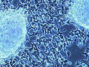
SH-SY5Y

SH-SY5Y is a human derived cell line used in scientific research. The original cell line, called SK-N-SH, from which it was subcloned was isolated from a bone marrow biopsy taken from a four-year-old female with neuroblastoma. SH-SY5Y cells are often used as in vitro models of neuronal function and differentiation. They are adrenergic in phenotype but also express dopaminergic markers and, as such, have been used to study Parkinson's Disease.
History
SH-SY5Y was cloned from a bone marrow biopsy derived line called SK-N-SH and first reported in 1973.[1] A neuroblast-like subclone of SK-N-SH, named SH-SY, was subcloned as SH-SY5, which was subcloned a third time to produce the SH-SY5Y line, first described in 1978.[2] The cloning process is essentially artificial selection, involving expansion of individual cells or a small group of cells that express a particular phenotype of interest, in this particular case neuron-like characteristic. The SH-SY5Y line is genetically female with two X and no Y chromosome, as expected given an origin from a four-year-old female.
Morphology
The cells typically grow in tissue culture in two distinct ways. Some grow into clumps of cells which float in the media, while others form clumps which stick to the dish. SH-SY5Y cells can spontaneously interconvert between these two phenotypes in vitro although the mechanisms underlying this process are not understood. However, the cell line is appreciated to be N-type (neuronal), given its morphology and the ability to differentiate the cells into along the neuronal lineage (in contrast to the S-type SH-EP subcloned cell line, also derived from SK-N-SH).[3] Cells with short spiny neurite-like processes migrate out from these adherent clumps. SH-SY5Y cells possess an abnormal chromosome 1, where there is an additional copy of a 1q segment and is referred to trisomy 1q. SH-SY5Y cells are known to be dopamine beta hydroxylase active, acetylcholinergic, glutamatergic and adenosinergic. The cells have very different growth phases, outlined in the surrounding pictures. The cells both propagate via mitosis and differentiate by extending neurites to the surrounding area. While dividing, the aggregated cells can look so different from the differentiated cells following the K'anul-index tumor cellularity (TNF, for Tumor Necrosis Factor), that new scientists often mistake one or the other for contamination. The dividing cells can form clusters of cells which are reminders of their cancerous nature, but certain treatments such as retinoic acid, BDNF, or TPA can force the cells to dendrify and differentiate. Moreover, induction by retinoic acid results in inhibition of cell growth and enhanced production of noradrenaline from SH-SY5Y cells.

Media and Cultivation
The most common growing cocktail used is a 1:1 mixture of DMEM and Ham's F12 medium and 10% supplemental fetal bovine serum. The DMEM usually contains 1.5 g/L sodium bicarbonate, 2 mM L-Glutamine, 1 mM sodium pyruvate and 0.1 mM nonessential amino acids. The cells are always grown at 37 degrees Celsius with 95% air and 5% carbon dioxide. It is advised to cultivate the cells in flasks which are coated for cell culture adhesion, this will aid in differention and dendrification of the neuroblastoma. In general the cells are quite robust and will grow in most widely used tissue culture media.
SH-SY5Y has a dopamine-β-hydroxylase activity and can convert glutamate to GABA. It will also form tumors in nude mice in around 3–4 weeks. The loss of neuronal characteristics has been described with increasing passage numbers. Therefore it is recommended not to be used after passage 20 or verify specific characteristics such as noradrenalin uptake or neuronal tumor markers.
Related links
- ATCC- American Type Culture Collection, a source of SH-SY5Y cells and other biologicials
- ATTC product data for SH-SY5Y cells, catalog CRL-2266
- Sigma Product Page
- Canals M, Angulo E, Casadó V, et al. (January 2005). "Molecular mechanisms involved in the adenosine A and A receptor-induced neuronal differentiation in neuroblastoma cells and striatal primary cultures". J. Neurochem. 92 (2): 337–48. doi:10.1111/j.1471-4159.2004.02856.x. PMID 15663481.
- Hillion J, Canals M, Torvinen M, et al. (May 2002). "Coaggregation, cointernalization, and codesensitization of adenosine A2A receptors and dopamine D2 receptors". J. Biol. Chem. 277 (20): 18091–7. doi:10.1074/jbc.M107731200. PMID 11872740.
- Okuda K, Kotake Y, Ohta S (July 2006). "Parkinsonism-preventing activity of 1-methyl-1,2,3,4-tetrahydroisoquinoline derivatives in C57BL mouse in vivo". Biol. Pharm. Bull. 29 (7): 1401–3. doi:10.1248/bpb.29.1401. PMID 16819177.
References
- ↑ Biedler JL, Helson L, Spengler BA (November 1973). "Morphology and growth, tumorigenicity, and cytogenetics of human neuroblastoma cells in continuous culture". Cancer Res. 33 (11): 2643–52. PMID 4748425.
- ↑ Biedler JL, Roffler-Tarlov S, Schachner M, Freedman LS (November 1978). "Multiple neurotransmitter synthesis by human neuroblastoma cell lines and clones". Cancer Res. 38 (11 Pt 1): 3751–7. PMID 29704.
- ↑ La Quaglia Michael P., Manchester Karen M. (1996). "A comparative analysis of neuroblastic and substrate-adherent human neuroblastoma cell lines". Journal of Pediatric Surgery 31 (2): 315–318. doi:10.1016/S0022-3468(96)90025-1.