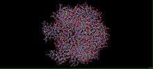
Polyomavirus capsid protein (VP1)
Viral Protein 1 (VP1), also known as Major Capsid Protein VP1, is the main component of the polyomavirus capsid. 360 VP1 molecules are organized in 72 pentamers to form the capsid. This protein also contains large unique regions that might be responsible for the characteristic properties observed in the Reoviridae family (such as rotavirus). Polyomaviruses have from five to seven proteins, including three capsid proteins, VP1, VP2, and VP3, and two proteins, T and t, which are involved in DNA replication.

History
Dr. Sarah Stewart and Dr. Bernice E. Eddy were the first ones to describe the polyoma virus. "Polyoma" is a word derived from the Greek roots "poly-", meaning many or much, and "-oma", meaning tumors. Until 2000 d.C, polyomaviruses were taxonomically classified as a genus in the family papovaviridae. With the publication of the Seventh Report of the International Committee on Taxonomy of Viruses, papovaviridae was split into two new families, papillomaviridae and polyomaviridae. Here we see some differences between them.[1]
| Polyomavirus | Papillomavirus | |
|---|---|---|
| Genome |
|
|
| Morphology |
|
|
| Pathogenesis | Most are asymptomatic, although can be oncogenic in hamsters | Cause various types of warts, epidermodysplasia verruciformis |
| Representative Viruses |
JC Virus
BK Virus
SV40 (Simian Virus 40)
|
At least 62 strains of Human Papillomaviruses
|
The major polyoma virus capsid protein,VP1, has been cloned in Escherichia coli. Under the inducible control of the hybrid tac promoter, VP1 constituted between 2 and 3% of the total host cell protein. The expressed VP, was purified to near homogeneity with initial yields to 10%. Optimal expression was temperature-dependent, and significant intracellular degradation could be demonstrated. The final product was obtained as one predominant isoelectric focusing species, without the pattern of post-translational modification seen in virus-infected eukaryotic cells. The purified VP1 from E. coli will be useful as a substrate for the purification of VP1 modification enzymes and in the study of inter-VP1 oligomerization.
Structure
This protein forms an icosahedral capsid with a T=7 symmetry and a 40 nm diameter. The capsid is composed of 72 pentamers linked to each other by disulfide bonds and associated with VP2 or VP3 proteins. Therefore, its subunit structure is a homomultimer; disulfide-linked. The VP1 can also interact with host 5HT2AR. Polyomavirus major capsid protein VP1 forms stable pentamers in low-ionic strength, neutral, or alkaline solutions.[3]

Electron microscopy showed that the pentamers, which correspond to viral capsomeres, can be self-assembled into a variety of polymorphic aggregates by lowering the pH, adding calcium, or raising the ionic strength. Some of the aggregates resembled the 500-A-diameter virus capsid, whereas other considerably larger or smaller capsids were also produced. The particular structures formed on transition to an environment favoring assembly depended on the pathway of the solvent changes as well as on the final conditions. Mass measurements from cryoelectron micrographs and image analysis of negatively stained specimens established that a distinctive 320-A-diameter particle consists of 24 close-packed pentamers arranged with octahedral symmetry. Comparison of this unexpected octahedral assembly with a 12-capsomere icosahedral aggregate and the 72-capsomere icosahedral virus capsid by computer graphics methods indicates that similar connections are made among trimers of pentamers in these shells of different size. The polymorphism in the assembly of VP1 pentamers can be related to the switching in bonding specificity required to build the virus capsid.
Composition of this protein
The sequence length of VP1 is 354 amino acids, being a complete sequence status. We should highlight the fact that this sequence is directly related to the polyomaviruses coat protein .
Functions
The main functions of the VP1 protein are to provide virion attachment to the target cell and to initiate the synthesis of the viral genome for the virus replication.

To provide the attachment to the target cell, the VP1 protein interacts with a N-linked glycoprotein containing terminal alpha(2-6)-linked sialic acids on the cell surface. Once attached, the virions enter predominantly by a ligand-inducible clathrin-dependent pathway and traffic to the Endoplasmic Reticulum. VP1 interpentamer disulfide bonds are isomarized in the Endoplasmatic Reticulum to activate initial uncoating. To enter to the nucleus of the cell, the importin recognition of VP2/Vp3 nuclear localization signals is essential. When the viral DNA finally enters into the nucleus, the replication of the genetic material begins and new viral DNA is created. In late phase of infection, neo-synthesized VP1 encapsulates replicated genomic DNA at nuclear domains called promyelocytic leukemia (PML) bodies, and participates in rearranging nucleosomes around the viral DNA.[4]
Once the synthesis of the viral genome is finished, the guanylic VP1 molecule used as the initiator of the process remains attached to the new RNA by a covalent bond with the guanines of the 5’ end of the new chain. To be used again the VP1 has to get free, what requires that the vp1-genome bond has to break. This break must be done in the phosphodiester bond between the hydroxyl group of the guanalilazed residue of the VP1 and the GMP of the 5’ of the genome, so the chain doesn’t get shorter in every replication.[5]
References
- ↑ Polyomaviridae
- ↑ Biology
- ↑ Major capsid protein VP1 - JC polyomavirus (JCPyV)
- ↑ Pho M.T., Ashok A., Atwood W.J.(2000)."JC virus enters human glial cells by clathrin-dependent receptor-mediated endocytosis."
- ↑ Garriga i Rigau, Damià (2009)."Anàlisi estructural de partícules i proteïnes del virus de la burcitis infecciosa"pag.92-94
Bibliography
http://www.uniprot.org/taxonomy/10632 http://jnci.oxfordjournals.org/content/94/4/267/F1/graphic-1.large.jpg http://www.microbiologybytes.com/virology/Polyomaviruses.html http://www.stanford.edu/group/virus/polyoma/2004temple/polyoma.html http://www.jbc.org/content/260/23/12803.full.pdf http://www.uniprot.org/uniprot/P03089 http://www.abc.es/Media/201105/30/escherichia-coli-sintomas--644x362.jpg http://bioinformatica.uab.es/biocomputacio/treballs00-01/rodriguez-rotllant/biologia_molecular.htm http://www.ncbi.nlm.nih.gov/pmc/articles/PMC1280588/ http://www.nature.com/emboj/journal/v17/n12/full/7591028a.html http://www.uniprot.org/uniprot/P03089