NDUFA5
| NADH dehydrogenase (ubiquinone) 1 alpha subcomplex, 5 | |||||||||||||
|---|---|---|---|---|---|---|---|---|---|---|---|---|---|
| Identifiers | |||||||||||||
| Symbols | NDUFA5 ; B13; CI-13KD-B; CI-13kB; NUFM; UQOR13 | ||||||||||||
| External IDs | OMIM: 601677 MGI: 1915452 HomoloGene: 3664 GeneCards: NDUFA5 Gene | ||||||||||||
| EC number | 1.6.5.3, 1.6.99.3 | ||||||||||||
| |||||||||||||
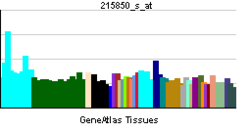
| RNA expression pattern | |||||||||||||
 | |||||||||||||
 | |||||||||||||
| More reference expression data | |||||||||||||
| Orthologs | |||||||||||||
| Species | Human | Mouse | |||||||||||
| Entrez | 4698 | 68202 | |||||||||||
| Ensembl | ENSG00000128609 | ENSMUSG00000023089 | |||||||||||
| UniProt | Q16718 | Q9CPP6 | |||||||||||
| RefSeq (mRNA) | NM_001282419 | NM_026614 | |||||||||||
| RefSeq (protein) | NP_001269348 | NP_080890 | |||||||||||
| Location (UCSC) |
Chr 7: 123.54 – 123.56 Mb |
Chr 6: 24.52 – 24.53 Mb | |||||||||||
| PubMed search | |||||||||||||
NADH dehydrogenase [ubiquinone] 1 alpha subcomplex subunit 5 is an enzyme that in humans is encoded by the NDUFA5 gene.[1] The NDUFA5 protein is a subunit of NADH dehydrogenase (ubiquinone), which is located in the mitochondrial inner membrane and is the largest of the five complexes of the electron transport chain.[2]
Structure
The NDUFA5 gene is located on the q arm of chromosome 7 and it spans 64,655 base pairs.[1] The gene produces a 13.5 kDa protein composed of 116 amino acids.[3][4] NDUFA5 is a subunit of the enzyme NADH dehydrogenase (ubiquinone), the largest of the respiratory complexes. The structure is L-shaped with a long, hydrophobic transmembrane domain and a hydrophilic domain for the peripheral arm that includes all the known redox centers and the NADH binding site.[2] It has been noted that the N-terminal hydrophobic domain has the potential to be folded into an alpha helix spanning the inner mitochondrial membrane with a C-terminal hydrophilic domain interacting with globular subunits of Complex I. The highly conserved two-domain structure suggests that this feature is critical for the protein function and that the hydrophobic domain acts as an anchor for the NADH dehydrogenase (ubiquinone) complex at the inner mitochondrial membrane. NDUFA5 is one of about 31 hydrophobic subunits that form the transmembrane region of Complex I. The protein localizes to the inner mitochondrial membrane as part of the 7 component-containing, water-soluble iron-sulfur protein (IP) fraction of complex I, although its specific role is unknown. It is assumed to undergo post-translational removal of the initiator methionine and N-acetylation of the next amino acid. The predicted secondary structure is primarily alpha helix, but the carboxy-terminal half of the protein has high potential to adopt a coiled-coil form. The amino-terminal part contains a putative beta sheet rich in hydrophobic amino acids that may serve as mitochondrial import signal. Related pseudogenes have also been identified on four other chromosomes.[1][5]
Function
The human NDUFA5 gene codes for the B13 subunit of complex I of the respiratory chain, which transfers electrons from NADH to ubiquinone. The NDUFA5 protein localizes to the mitochondrial inner membrane and it is thought to aid in this transfer of electrons.[1] Initially, NADH binds to Complex I and transfers two electrons to the isoalloxazine ring of the flavin mononucleotide (FMN) prosthetic arm to form FMNH2. The electrons are transferred through a series of iron-sulfur (Fe-S) clusters in the prosthetic arm and finally to coenzyme Q10 (CoQ), which is reduced to ubiquinol (CoQH2). The flow of electrons changes the redox state of the protein, resulting in a conformational change and pK shift of the ionizable side chain, which pumps four hydrogen ions out of the mitochondrial matrix.[2] The high degree of conservation of NDUFA5 extending to plants and fungi indicates its functional significance in the enzyme complex.[6]
Clinical significance
NDUFA5, ATP5A1 and ATP5A1 all show consistently reduced expression in brains of autism patients. Mitochondrial dysfunction and impaired ATP synthesis can result in oxidative stress, which may play a role in the development of autism.[7][8]
Interactions
NDUFA5 has many protein-protein interactions, such as ubiquitin C and with members of the NADH dehydrogenase [ubiquinone] 1 beta subcomplex, including NDUFB1, NDUFB9 and NDUFB10.[1]
References
- 1 2 3 4 5 "Entrez Gene: NDUFA5 NADH dehydrogenase (ubiquinone) 1 alpha subcomplex, 5".
- 1 2 3 Pratt, Donald Voet, Judith G. Voet, Charlotte W. (2013). "18". Fundamentals of biochemistry : life at the molecular level (4th ed.). Hoboken, NJ: Wiley. pp. 581–620. ISBN 9780470547847.
- ↑ Zong NC, Li H, Li H, Lam MP, Jimenez RC, Kim CS, Deng N, Kim AK, Choi JH, Zelaya I, Liem D, Meyer D, Odeberg J, Fang C, Lu HJ, Xu T, Weiss J, Duan H, Uhlen M, Yates JR, Apweiler R, Ge J, Hermjakob H, Ping P (Oct 2013). "Integration of cardiac proteome biology and medicine by a specialized knowledgebase". Circulation Research 113 (9): 1043–53. doi:10.1161/CIRCRESAHA.113.301151. PMC 4076475. PMID 23965338.
- ↑ "NDUFA5 - NADH dehydrogenase [ubiquinone] 1 alpha subcomplex subunit 5". Cardiac Organellar Protein Atlas Knowledgebase (COPaKB).
- ↑ Emahazion T, Beskow A, Gyllensten U, Brookes AJ (Nov 1998). "Intron based radiation hybrid mapping of 15 complex I genes of the human electron transport chain". Cytogenet Cell Genet 82 (1-2): 115–9. doi:10.1159/000015082. PMID 9763677.
- ↑ Russell MW, du Manoir S, Collins FS, Brody LC (Mar 1997). "Cloning of the human NADH: ubiquinone oxidoreductase subunit B13: localization to chromosome 7q32 and identification of a pseudogene on 11p15". Mamm Genome 8 (1): 60–1. doi:10.1007/s003359900350. PMID 9021153.
- ↑ Anitha, A; Nakamura, K; Thanseem, I; Matsuzaki, H; Miyachi, T; Tsujii, M; Iwata, Y; Suzuki, K; Sugiyama, T; Mori, N (2013). "Downregulation of the expression of mitochondrial electron transport complex genes in autism brains". Brain Pathology 23 (3): 294–302. doi:10.1111/bpa.12002. PMID 23088660.
- ↑ Marui, T; Funatogawa, I; Koishi, S; Yamamoto, K; Matsumoto, H; Hashimoto, O; Jinde, S; Nishida, H; Sugiyama, T; Kasai, K; Watanabe, K; Kano, Y; Kato, N (2011). "The NADH-ubiquinone oxidoreductase 1 alpha subcomplex 5 (NDUFA5) gene variants are associated with autism". Acta Psychiatrica Scandinavica 123 (2): 118–24. doi:10.1111/j.1600-0447.2010.01600.x. PMID 20825370.
Further reading
- Smeitink J, van den Heuvel L (1999). "Human mitochondrial complex I in health and disease.". Am. J. Hum. Genet. 64 (6): 1505–10. doi:10.1086/302432. PMC 1377894. PMID 10330338.
- Pata I, Tensing K, Metspalu A (1997). "A human cDNA encoding the homologue of NADH: ubiquinone oxidoreductase subunit B13.". Biochim. Biophys. Acta 1350 (2): 115–8. doi:10.1016/s0167-4781(96)00208-4. PMID 9048877.
- "Toward a complete human genome sequence.". Genome Res. 8 (11): 1097–108. 1999. doi:10.1101/gr.8.11.1097. PMID 9847074.
- Loeffen JL, Triepels RH, van den Heuvel LP, et al. (1999). "cDNA of eight nuclear encoded subunits of NADH:ubiquinone oxidoreductase: human complex I cDNA characterization completed.". Biochem. Biophys. Res. Commun. 253 (2): 415–22. doi:10.1006/bbrc.1998.9786. PMID 9878551.
- Tensing K, Pata I, Wittig I, et al. (1999). "Genomic organization of the human complex I 13-kDa subunit gene NDUFA5.". Cytogenet. Cell Genet. 84 (1-2): 125–7. doi:10.1159/000015237. PMID 10343126.
- Strausberg RL, Feingold EA, Grouse LH, et al. (2003). "Generation and initial analysis of more than 15,000 full-length human and mouse cDNA sequences.". Proc. Natl. Acad. Sci. U.S.A. 99 (26): 16899–903. doi:10.1073/pnas.242603899. PMC 139241. PMID 12477932.
- Hillier LW, Fulton RS, Fulton LA, et al. (2003). "The DNA sequence of human chromosome 7.". Nature 424 (6945): 157–64. doi:10.1038/nature01782. PMID 12853948.
- Ota T, Suzuki Y, Nishikawa T, et al. (2004). "Complete sequencing and characterization of 21,243 full-length human cDNAs.". Nat. Genet. 36 (1): 40–5. doi:10.1038/ng1285. PMID 14702039.
- Gerhard DS, Wagner L, Feingold EA, et al. (2004). "The status, quality, and expansion of the NIH full-length cDNA project: the Mammalian Gene Collection (MGC).". Genome Res. 14 (10B): 2121–7. doi:10.1101/gr.2596504. PMC 528928. PMID 15489334.
- Rual JF, Venkatesan K, Hao T, et al. (2005). "Towards a proteome-scale map of the human protein-protein interaction network.". Nature 437 (7062): 1173–8. doi:10.1038/nature04209. PMID 16189514.
This article incorporates text from the United States National Library of Medicine, which is in the public domain.