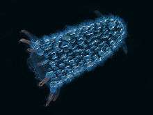
Bioluminescence
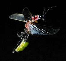

Bioluminescence is the production and emission of light by a living organism. It is a form of chemiluminescence. Bioluminescence occurs widely in marine vertebrates and invertebrates, as well as in some fungi, microorganisms including some bioluminescent bacteria and terrestrial invertebrates such as fireflies. In some animals, the light is produced by symbiotic organisms such as Vibrio bacteria.
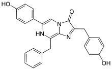
The principal chemical reaction in bioluminescence involves the light-emitting pigment luciferin and the enzyme luciferase, assisted by other proteins such as aequorin in some species. The enzyme catalyzes the oxidation of luciferin. In some species, the type of luciferin requires cofactors such as calcium or magnesium ions, and sometimes also the energy-carrying molecule adenosine triphosphate (ATP). In evolution, luciferins vary little: one in particular, coelenterazine, is found in nine different animal (phyla), though in some of these, the animals obtain it through their diet. Conversely, luciferases vary widely in different species. Bioluminescence has arisen over forty times in evolutionary history.
Both Aristotle and Pliny the Elder mentioned that damp wood sometimes gives off a glow and many centuries later Robert Boyle showed that oxygen was involved in the process, both in wood and in glow-worms. It was not until the late nineteenth century that bioluminescence was properly investigated. The phenomenon is widely distributed among animal groups, especially in marine environments where dinoflagellates cause phosphorescence in the surface layers of water. On land it occurs in fungi, bacteria and some groups of invertebrates, including insects.
The uses of bioluminescence by animals include counter-illumination camouflage, mimicry of other animals, for example to lure prey, and signalling to other individuals of the same species, such as to attract mates. In the laboratory, luciferase-based systems are used in genetic engineering and for biomedical research. Other researchers are investigating the possibility of using bioluminescent systems for street and decorative lighting, and a bioluminescent plant has been created.[1]
History
Before the development of the safety lamp for use in coal mines, dried fish skins were used in Britain and Europe as a weak source of light.[2] This experimental form of illumination avoided the necessity of using candles which risked sparking explosions of firedamp.[3] Another safe source of illumination in mines was bottles containing fireflies.[4] In 1920, the American zoologist E. Newton Harvey published a monograph, The Nature of Animal Light, summarizing early work on bioluminescence. Harvey notes that Aristotle mentions light produced by dead fish and flesh, and that both Aristotle and Pliny the Elder (in his Natural History) mention light from damp wood. He also records that Robert Boyle experimented on these light sources, and showed that both they and the glow-worm require air for light to be produced. Harvey notes that in 1753, J. Baker identified the flagellate Noctiluca "as a luminous animal" "just visible to the naked eye",[5] and in 1854 Johann Florian Heller (1813-1871) identified strands (hyphae) of fungi as the source of light in dead wood.[6]
Tuckey, in his posthumous 1818 Narrative of the Expedition to the Zaire, described catching the animals responsible for luminescence. He mentions pellucids, crustaceans (to which he ascribes the milky whiteness of the water), and cancers (shrimps and crabs). Under the microscope he described the "luminous property" to be in the brain, resembling "a most brilliant amethyst about the size of a large pin's head".[7]
Charles Darwin noticed bioluminescence in the sea, describing it in his Journal:
While sailing in these latitudes on one very dark night, the sea presented a wonderful and most beautiful spectacle. There was a fresh breeze, and every part of the surface, which during the day is seen as foam, now glowed with a pale light. The vessel drove before her bows two billows of liquid phosphorus, and in her wake she was followed by a milky train. As far as the eye reached, the crest of every wave was bright, and the sky above the horizon, from the reflected glare of these livid flames, was not so utterly obscure, as over the rest of the heavens.[8]
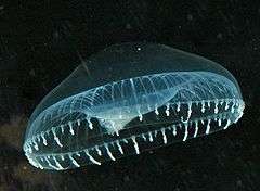
Darwin also observed a luminous "jelly-fish of the genus Dianaea"[8] and noted that "When the waves scintillate with bright green sparks, I believe it is generally owing to minute crustacea. But there can be no doubt that very many other pelagic animals, when alive, are phosphorescent."[8] He guessed that "a disturbed electrical condition of the atmosphere"[8] was probably responsible. Daniel Pauly comments that Darwin "was lucky with most of his guesses, but not here",[9] noting that biochemistry was too little known, and that the complex evolution of the marine animals involved "would have been too much for comfort".[9]
Bioluminescence attracted the attention of the United States Navy in the Cold War, since submarines in some waters can create a bright enough wake to be detected; a German submarine was sunk in the First World War, having been detected in this way. The navy was interested in predicting when such detection would be possible, and hence guiding their own submarines to avoid detection.[11]
Among the anecdotes of navigation by bioluminescence, the Apollo 13 astronaut Jim Lovell recounted how as a navy pilot he had found his way back to his aircraft carrier USS Shangri-La when his navigation systems failed. Turning off his cabin lights, he saw the glowing wake of the ship, and was able to fly to it and land safely.[12]
The French pharmacologist Raphaël Dubois carried out work on bioluminescence in the late nineteenth century. He studied click beetles (Pyrophorus) and the marine bivalve mollusc Pholas dactylus. He refuted the old idea that bioluminescence came from phosphorus,[13][lower-alpha 1] and demonstrated that the process was related to the oxidation of a specific compound, which he named luciferin, by an enzyme.[15] He sent Harvey siphons from the mollusc preserved in sugar. Harvey had become interested in bioluminescence as a result of visiting the South Pacific and Japan and observing phosphorescent organisms there. He studied the phenomenon for many years. His research aimed to demonstrate that luciferin, and the enzymes that act on it is to produce light, were interchangeable between species, showing that all bioluminescent organisms had a common ancestor. However, he found this hypothesis to be false, with different organisms having major differences in the composition of their light-producing proteins. He spent the next thirty years purifying and studying the components, but it fell to the young Japanese chemist Osamu Shimomura to be the first to obtain crystalline luciferin. He used the sea firefly Vargula hilgendorfii, but it was another ten years before he discovered the chemical's structure and was able to publish his 1957 paper Crystalline Cypridina Luciferin.[16] More recently, Martin Chalfie, Osamu Shimomura and Roger Y. Tsien won the 2008 Nobel Prize in Chemistry for their 1961 discovery and development of green fluorescent protein as a tool for biological research.[17]
Chemical mechanism
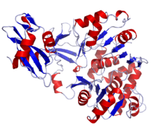
Bioluminescence is a form of chemiluminescence where light energy is released by a chemical reaction. Fireflies, anglerfish, and other organisms produce the light-emitting pigment luciferin and the enzyme luciferase.[18] Luciferin reacts with oxygen to create light:
Carbon dioxide (CO2), adenosine monophosphate (AMP) and phosphate groups (PP) are released as waste products. Luciferase catalyzes the reaction, which may be mediated by cofactors such as calcium (Ca2+) or magnesium (Mg2+) ions, and for some types of luciferin (L) also the energy-carrying molecule adenosine triphosphate (ATP). The reaction can occur either inside or outside the cell. In bacteria such as Vibrio, the expression of genes related to bioluminescence is controlled by the lux operon.[19]
In evolution, luciferins generally vary little: one in particular, coelenterazine, is the light emitting pigment for nine ancient phyla (groups of very different organisms), including polycystine radiolaria, Cercozoa (Phaeodaria), protozoa, comb jellies, cnidaria including jellyfish and corals, crustaceans, molluscs, arrow worms and vertebrates (ray-finned fish). Not all these organisms synthesize coelenterazine: some of them obtain it through their diet.[18] Conversely, luciferase enzymes vary widely and tend to be different in each species. Overall, bioluminescence has arisen over forty times in evolutionary history.[18]
Luciferin-luciferase reactions are not the only way that organisms produce light. The parchment worm Chaetopterus (a marine Polychaete) makes use of another photoprotein, aequorin, instead of luciferase. When calcium ions are added, the aequorin's rapid catalysis creates a brief flash quite unlike the prolonged glow produced by luciferase. In a second, much slower, step luciferin is regenerated from the oxidised (oxyluciferin) form, allowing it to recombine with aequorin, in readiness for a subsequent flash. Photoproteins are thus enzymes, but with unusual reaction kinetics.[20]
In the hydrozoan jellyfish Aequorea victoria,[21] some of the blue light released by aequorin in contact with calcium ions is absorbed by green fluorescent protein; it in turn releases green light.[22]
Distribution
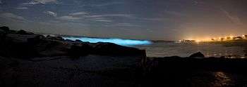
Bioluminescence occurs widely among animals, especially in the open sea, including fish, jellyfish, comb jellies, crustaceans, and cephalopod molluscs; in some fungi and bacteria; and in various terrestrial invertebrates including insects. Many, perhaps most deep-sea animals produce light. Most marine light-emission is in the blue and green light spectrum. However, some loose-jawed fish emit red and infrared light, and the genus Tomopteris emits yellow light.[18][23]
The most frequently encountered bioluminescent organisms may be the dinoflagellates present in the surface layers of the sea, which are responsible for the sparkling phosphorescence sometimes seen at night in disturbed water. At least eighteen genera exhibit luminosity.[18] A different effect is the thousands of square miles of the ocean which shine with the light produced by bioluminescent bacteria, known as mareel or the milky seas effect.[24]
Non-marine bioluminescence is less widely distributed, the two best-known cases being in fireflies and glow worms. Other invertebrates including insect larvae, annelids and arachnids possess bioluminescent abilities. Some forms of bioluminescence are brighter (or exist only) at night, following a circadian rhythm.
Uses in nature
Bioluminescence has several functions in different taxa. Haddock et al. (2010) list as more or less definite functions in marine organisms the following: defensive functions of startle, counterillumination (camouflage), misdirection (smoke screen), distractive body parts, burglar alarm (making predators easier for higher predators to see), and warning to deter settlers; offensive functions of lure, stun or confuse prey, illuminate prey, and mate attraction/recognition. It is much easier for researchers to detect that a species is able to produce light than to analyse the chemical mechanisms or to prove what function the light serves.[18] In some cases the function is unknown, as with species in three families of earthworm (Oligochaeta), such as Diplocardia longa where the coelomic fluid produces light when the animal moves.[25] The following functions are reasonably well established in the named organisms.
Counterillumination camouflage
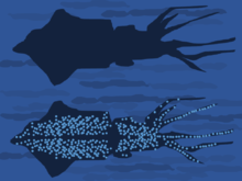
In many animals of the deep sea, including several squid species, bacterial bioluminescence is used for camouflage by counterillumination, in which the animal matches the overhead environmental light as seen from below.[26] In these animals, photoreceptors control the illumination to match the brightness of the background.[26] These light organs are usually separate from the tissue containing the bioluminescent bacteria. However, in one species, Euprymna scolopes, the bacteria are an integral component of the animal's light organ.[27]
Attraction
A fungus gnat from New Zealand, Arachnocampa luminosa, lives in the predator-free environment of caves and its larvae emit bluish-green light. They dangle silken threads that glow and attract flying insects, and wind in their fishing-lines when prey becomes entangled.[28] The bioluminescence of the larvae of another fungus gnat from North America which lives on streambanks and under overhangs has a similar function. Orfelia fultoni builds sticky little webs and emits light of a deep blue colour. It has an inbuilt biological clock and, even when kept in total darkness, turns its light on and off in a circadian rhythm.[29]
Fireflies use light to attract mates. Two systems are involved according to species; in one, females emit light from their abdomens to attract males; in the other, flying males emit signals to which the sometimes sedentary females respond.[25][30] Click beetles emit an orange light from the abdomen when flying and a green light from the thorax when they are disturbed or moving about on the ground. The former is probably a sexual attractant but the latter may be defensive.[25] Larvae of the click beetle Pyrophorus nyctophanus live in the surface layers of termite mounds in Brazil. They light up the mounds by emitting a bright greenish glow which attracts the flying insects on which they feed.[25]
In the marine environment, use of luminescence for mate attraction is chiefly known among ostracods, small shrimplike crustaceans, especially in the Cyprinidae family. Pheromones may be used for long-distance communication, with bioluminescence used at close range to enable mates to "home in".[18] A polychaete worm, the Bermuda fireworm creates a brief display, a few nights after the full moon, when the female lights up to attract males.[31]
Defense
Many cephalopods, including at least 70 genera of squid, are bioluminescent.[18] Some squid and small crustaceans use bioluminescent chemical mixtures or bacterial slurries in the same way as many squid use ink. A cloud of luminescent material is expelled, distracting or repelling a potential predator, while the animal escapes to safety.[18] The deep sea squid Octopoteuthis deletron may autotomise portions of its arms which are luminous and continue to twitch and flash, thus distracting a predator while the animal flees.[18]
Dinoflagellates may use bioluminescence for defence against predators. They shine when they detect a predator, possibly making the predator itself more vulnerable by attracting the attention of predators from higher trophic levels.[18] Grazing copepods release any phytoplankton cells that flash, unharmed; if they were eaten they would make the copepods glow, attracting predators, so the phytoplankton's bioluminescence is defensive. The problem of shining stomach contents is solved (and the explanation corroborated) in predatory deep-sea fishes: their stomachs have a black lining able to keep the light from any bioluminescent fish prey which they have swallowed from attracting larger predators.[9]
The sea-firefly is a small crustacean living in sediment. At rest it emits a dull glow but when disturbed it darts away leaving a cloud of shimmering blue light to confuse the predator. During World War II it was gathered and dried for use by the Japanese military as a source of light during clandestine operations.[16]
The larvae of railroad worms (Phrixothrix) have paired photic organs on each body segment, able to glow with green light; these are thought to have a defensive purpose.[32] They also have organs on the head which produce red light; they are the only terrestrial organisms to emit light of this colour.[33]
Warning
Aposematism is a widely used function of bioluminescence, providing a warning that the creature concerned is unpalatable. It is suggested that many firefly larvae glow to repel predators; millipedes glow for the same purpose.[34] Some marine organisms are believed to emit light for a similar reason. These include scale worms, jellyfish and brittle stars but further research is needed to fully establish the function of the luminescence. Such a mechanism would be of particular advantage to soft-bodied cnidarians if they were able to deter predation in this way.[18] The limpet Latia neritoides is the only known freshwater gastropod that emits light. It produces greenish luminescent mucus which may have an anti-predator function.[35] The marine snail Hinea brasiliana uses flashes of light, probably to deter predators. The blue-green light is emitted through the translucent shell, which functions as an efficient diffuser of light.[36]
Communication
Communication in the form of quorum sensing plays a role in the regulation of luminescence in many species of bacteria. Small extracellularly secreted molecules stimulate the bacteria to turn on genes for light production when cell density, measured by concentration of the secreted molecules, is high.[18]
Pyrosomes are colonial tunicates and each zooid has a pair of luminescent organs on either side of the inlet siphon. When stimulated by light, these turn on and off, causing rhythmic flashing. No neural pathway runs between the zooids, but each responds to the light produced by other individuals, and even to light from other nearby colonies.[37] Communication by light emission between the zooids enables coordination of colony effort, for example in swimming where each zooid provides part of the propulsive force.[38]
Some bioluminous bacteria infect nematodes that parasitize Lepidoptera larvae. When these caterpillars die, their luminosity may attract predators to the dead insect thus assisting in the dispersal of both bacteria and nematodes.[25] A similar reason may account for the many species of fungi that emit light. Species in the genera Armillaria, Mycena, Omphalotus, Panellus, Pleurotus and others do this, emitting usually greenish light from the mycelium, cap and gills. This may attract night-flying insects and aid in spore dispersal, but other functions may also be involved.[25]
Quantula striata is the only known bioluminescent terrestrial mollusc. Pulses of light are emitted from a gland near the front of the foot and may have a communicative function, although the adaptive significance is not fully understood.[39]
Mimicry

Bioluminescence is used by a variety of animals to mimic other species. Many species of deep sea fish such as the anglerfish and dragonfish make use of aggressive mimicry to attract prey. They have an appendage on their heads called an esca that contains bioluminescent bacteria able to produce a long-lasting glow which the fish can control. The glowing esca is dangled or waved about to lure small animals to within striking distance of the fish.[18][40]
The cookiecutter shark uses bioluminescence to camouflage its underside by counterillumination, but a small patch near its pectoral fins remains dark, appearing as a small fish to large predatory fish like tuna and mackerel swimming beneath it. When such fish approach the lure, they are bitten by the shark.[41][42]
Female Photuris fireflies sometimes mimic the light pattern of another firefly, Photinus, to attract its males as prey. In this way they obtain both food and the defensive chemicals named lucibufagins, which Photuris cannot synthesize.[43]
South American giant cockroaches of the genus Lucihormetica were believed to be the first known example of defensive mimicry, emitting light in imitation of bioluminescent, poisonous click beetles.[44] However, doubt has been cast on this assertion, and there is no conclusive evidence that the cockroaches are bioluminescent.[45][46]
Illumination
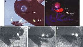
While most marine bioluminescence is green to blue, some deep sea barbeled dragonfishes in the genera Aristostomias, Pachystomias and Malacosteus emit a red glow. This adaptation allows the fish to see red-pigmented prey, which are normally invisible in the deep ocean environment where red light has been filtered out by the water column.[47]
The black dragonfish (also called the northern stoplight loosejaw) Malacosteus niger is believed to be one of the only fish to produce a red glow. Its eyes, however, are insensitive to this wavelength; it has an additional retinal pigment which fluoresces blue-green when illuminated. This alerts the fish to the presence of its prey. The additional pigment is thought to be assimilated from chlorophyll derivatives found in the copepods which form part of its diet.[48]
Biotechnology
Biology and medicine
Bioluminescent organisms are a target for many areas of research. Luciferase systems are widely used in genetic engineering as reporter genes, each producing a different colour by fluorescence,[49][50] and for biomedical research using bioluminescence imaging.[51][52][53] For example, the firefly luciferase gene was used as early as 1986 for research using transgenic tobacco plants.[54] Vibrio bacteria symbiose with marine invertebrates such as the Hawaiian bobtail squid (Euprymna scolopes), are key experimental models for bioluminescence.[55][56] Bioluminescent activated destruction is an experimental cancer treatment.[57] See also optogenetics which involves the use of light to control cells in living tissue, typically neurons, that have been genetically modified to express light-sensitive ion channels, and also see biophoton, a photon of non-thermal origin in the visible and ultraviolet spectrum emitted from a biological system.
Light production
The structures of photophores, the light producing organs in bioluminescent organisms, are being investigated by industrial designers. Engineered bioluminescence could perhaps one day be used to reduce the need for street lighting, or for decorative purposes if it becomes possible to produce light that is both bright enough and can be sustained for long periods at a workable price.[11][58][59] The gene that makes the tails of fireflies glow has been added to mustard plants. The plants glow faintly for an hour when touched, but a sensitive camera is needed to see the glow.[60] University of Wisconsin–Madison is researching the use of genetically engineered bioluminescent E. coli bacteria, for use as bioluminescent bacteria in a light bulb.[61] In June 2013 the Glowing Plant project raised nearly $500,000 on the crowdfunding site Kickstarter to create a bioluminescent plant.[62] An iGEM team from Cambridge (England) has started to address the problem that luciferin is consumed in the light-producing reaction by developing a genetic biotechnology part that codes for a luciferin regenerating enzyme from the North American firefly; this enzyme "helps to strengthen and sustain light output".[63]
Notes
References
- ↑ Callaway, E. 2013. Glowing plants spark debate. Nature, 498:15-16, 04 June 2013. http://www.nature.com/news/glowing-plants-spark-debate-1.13131
- ↑ Smiles, Samuel (1862). Lives of the Engineers. Volume III (George and Robert Stephenson). London: John Murray. p. 107. ISBN 0-7153-4281-9. (ISBN refers to the David & Charles reprint of 1968 with an introduction by L. T. C. Rolt)
- ↑ Freese, Barbara (2006). Coal: A Human History. Arrow. p. 51. ISBN 978-0-09-947884-3.
- ↑ Fordyce, William (20 July 1973). A history of coal, coke and coal fields and the manufacture of iron in the North of England. Graham.
- ↑ Harvey cites this as Baker, J.: 1743-1753, The Microscope Made Easy and Employment for the Microscope.
- ↑ Harvey, E. Newton (1920). The Nature of Animal Light. Philadelphia & London: J. B. Lippencott. Page 1.
- ↑ Tuckey, James Hingston (May 1818). Thomson, Thomas, ed. Narrative of the Expedition to the Zaire. Annals of Philosophy. volume XI. p. 392. Retrieved 22 April 2015.
- 1 2 3 4 Darwin, Charles (1839). Narrative of the surveying voyages of His Majesty's Ships Adventure and Beagle between the years 1826 and 1836, describing their examination of the southern shores of South America, and the Beagle's circumnavigation of the globe. Journal and remarks. 1832-1836. Henry Colburn. pp. 190–192.
- 1 2 3 Pauly, Daniel (13 May 2004). Darwin's Fishes: An Encyclopedia of Ichthyology, Ecology, and Evolution. Cambridge University Press. pp. 15–16. ISBN 978-1-139-45181-9.
- ↑ Shimomura O. (August 1995). "A short story of aequorin". The Biological bulletin 189 (1): 1–5. doi:10.2307/1542194. JSTOR 1542194. PMID 7654844.
- 1 2 "How illuminating". The Economist. 10 March 2011. Retrieved 6 December 2014.
- ↑ Huth, John Edward (15 May 2013). The Lost Art of Finding Our Way. Harvard University Press. p. 423. ISBN 978-0-674-07282-4.
- ↑ Reshetiloff, Kathy (1 July 2001). "Chesapeake Bay night-lights add sparkle to woods, water". Bay Journal. Retrieved 16 December 2014.
- ↑ "Luminescence". Encyclopaedia Britannica. Retrieved 16 December 2014.
- ↑ Poisson, Jacques (April 2010). "Raphaël Dubois, from pharmacy to bioluminescence". Rev Hist Pharm (Paris) (in French) (France) 58 (365): 51–56. ISSN 0035-2349. PMID 20533808.
- 1 2 Pieribone, Vincent; Gruber, David F. (2005). Aglow in the Dark: The Revolutionary Science of Biofluorescence. Harvard University Press. pp. 35–41. ISBN 978-0-674-01921-8.
- ↑ "The Nobel Prize in Chemistry 2008". 8 October 2008. Retrieved 23 November 2014.
- 1 2 3 4 5 6 7 8 9 10 11 12 13 14 Haddock, Steven H.D.; Moline, Mark A.; Case, James F. (2010). "Bioluminescence in the Sea". Annual Review of Marine Science 2: 443–493. Bibcode:2010ARMS....2..443H. doi:10.1146/annurev-marine-120308-081028. PMID 21141672.
- ↑ Hastings, J.W. (1983). "Biological diversity, chemical mechanisms, and the evolutionary origins of bioluminescent systems". J. Mol. Evol. 19 (5): 309–21. doi:10.1007/BF02101634. ISSN 1432-1432. PMID 6358519.
- ↑ Shimomura, O.; Johnson, F.H. (1975). "Regeneration of the photoprotein aequorin". Nature 256 (5514): 236–238. Bibcode:1975Natur.256..236S. doi:10.1038/256236a0. PMID 239351.
- ↑ Shimomura O, Johnson FH, Saiga Y; Johnson; Saiga (1962). "Extraction, purification and properties of aequorin, a bioluminescent protein from the luminous hydromedusan, Aequorea". J Cell Comp Physiol 59 (3): 223–39. doi:10.1002/jcp.1030590302. PMID 13911999.
- ↑ Morise, H.; Shimomura, O; Johnson, F.H.; Winant, J.; Shimomura; Johnson; Winant (1974). "Intermolecular energy transfer in the bioluminescent system of Aequorea". Biochemistry 13 (12): 2656–62. doi:10.1021/bi00709a028. PMID 4151620.
- ↑ Sparks, John S.; Schelly, Robert C.; Smith, W. Leo; Davis, Matthew P.; Tchernov, Dan; Pieribone, Vincent A.; Gruber, David F. (January 8, 2014). "The Covert World of Fish Biofluorescence: A Phylogenetically Widespread and Phenotypically Variable Phenomenon". PLoS ONE 9 (1): e83259. Bibcode:2014PLoSO...983259S. doi:10.1371/journal.pone.0083259. PMC 3885428. PMID 24421880.
- ↑ Ross, Alison (27 September 2005). "'Milky seas' detected from space". BBC. Retrieved 13 March 2013.
- 1 2 3 4 5 6 Viviani, Vadim (2009-02-17). "Terrestrial bioluminescence". Retrieved 2014-11-26.
- 1 2 Young, R.E.; Roper, C.F. (1976). "Bioluminescent countershading in midwater animals: evidence from living squid". Science 191 (4231): 1046–8. Bibcode:1976Sci...191.1046Y. doi:10.1126/science.1251214. PMID 1251214.
- ↑ Tong, D; Rozas, N.S.; Oakley, T.H.; Mitchell, J.; Colley, N.J.; McFall-Ngai, M.J. (2009). "Evidence for light perception in a bioluminescent organ". Proceedings of the National Academy of Sciences of the United States of America 106 (24): 9836–41. Bibcode:2009PNAS..106.9836T. doi:10.1073/pnas.0904571106. PMC 2700988. PMID 19509343.
- ↑ Broadley, R.; Stringer, I. (2009). "Larval behaviour of the New Zealand glowworm, Arachnocampa luminosa (Diptera: Keroplatidae), in bush and caves". Kerala: Research Signpost. Bioluminescence in focus: 325–355.
- ↑ Fulcher, Bob. "Lovely and Dangerous Lights" (PDF). Tennessee Conservationist Magazine. Retrieved 28 November 2014.
- ↑ Stanger-Hall, K.F.; Lloyd, J.E.; Hillis, D.M. (2007). "Phylogeny of North American fireflies (Coleoptera: Lampyridae): implications for the evolution of light signals". Molecular Phylogenetics and Evolution 45 (1): 33–49. doi:10.1016/j.ympev.2007.05.013. PMID 17644427.
- ↑ Shimomura, Osamu (2012). Bioluminescence: Chemical Principles and Methods. World Scientific. p. 234. ISBN 978-981-4366-08-3.
- ↑ Branham, Marc. "Glow-worms, railroad-worms (Insecta: Coleoptera: Phengodidae)". Featured Creatures. University of Florida. Retrieved 29 November 2014.
- ↑ Viviani, Vadim R.; Bechara, Etelvino J.H. (1997). "Bioluminescence and Biological Aspects of Brazilian Railroad-Worms (Coleoptera: Phengodidae)". Annals of the Entomological Society of America 90 (3): 389–398. doi:10.1093/aesa/90.3.389.
- ↑ Marek, Paul; Papaj, Daniel; Yeager, Justin; Molina, Sergio; Moore, Wendy. "Bioluminescent aposematism in millipedes". Current Biology 21 (18): R680–R681. doi:10.1016/j.cub.2011.08.012. PMC 3221455. PMID 21959150.
- ↑ Meyer-Rochow, V. B.; Moore, S. (1988). "Biology of Latia neritoides Gray 1850 (Gastropoda, Pulmonata, Basommatophora): the Only Light-producing Freshwater Snail in the World". Internationale Revue der gesamten Hydrobiologie und Hydrographie 73 (1): 21–42. doi:10.1002/iroh.19880730104.
- ↑ Deheyn, Dimitri D.; Wilson, Nerida G. (2010). "Bioluminescent signals spatially amplified by wavelength-specific diffusion through the shell of a marine snail". Proceedings of the Royal Society 278: 2112–2121. doi:10.1098/rspb.2010.2203.
- ↑ Bowlby, Mark R., Edith Widder, James Case (1990). "Patterns of stimulated bioluminescence in two pyrosomes (Tunicata: Pyrosomatidae)". Biological Bulletin 179 (3). JSTOR 1542326.
- ↑ Encyclopedia of the Aquatic World. Marshall Cavendish. January 2004. p. 1115. ISBN 978-0-7614-7418-0.
- ↑ Copeland J.; Daston, M. M. (1989). "Bioluminescence in the terrestrial snail Quantula (Dyakia) striata". Malacologia 30 (1–2): 317–324.
- ↑ Young, Richard Edward (October 1983). "Oceanic Bioluminescence: an Overview of General Functions". Bulletin of Marine Science 33 (4): 829–845.
- ↑ Martin, R. Aidan. "Biology of Sharks and Rays: Cookiecutter Shark". ReefQuest Centre for Shark Research. Retrieved 13 March 2013.
- ↑ Milius, S. (1 August 1998). "Glow-in-the-dark shark has killer smudge". Science News. Retrieved 13 March 2013.
- ↑ Thomas Eisner, Michael A. Goetz, David E. Hill, Scott R. Smedley, Jarrold Meinwald (1997). "Firefly "femmes fatales" acquire defensive steroids (lucibufagins) from their firefly prey". Proceedings of the National Academy of Sciences of the United States of America 94 (18): 9723–9728. Bibcode:1997PNAS...94.9723E. doi:10.1073/pnas.94.18.9723.
- ↑ Sullivan, Rachel. "Out of the darkness". ABC Science. Retrieved 17 December 2014.
- ↑ Greven, Hartmut; Zwanzig, Nadine (2013). "Courtship, Mating, and Organisation of the Pronotum in the Glowspot Cockroach Lucihormetica verrucosa (Brunner von Wattenwyl, 1865) (Blattodea: Blaberidae)". Entomologie heute 25: 77–97.
- ↑ Merritt, David J. (2013). "Standards of evidence for bioluminescence in cockroaches". Naturwissenschaften 100 (7): 697–698. Bibcode:2013NW....100..697M. doi:10.1007/s00114-013-1067-9.
- ↑ Douglas, R.H.; Mullineaux, C.W.; Partridge, J.C. (29 September 2000). "Long-wave sensitivity in deep-sea stomiid dragonfish with far-red bioluminescence: evidence for a dietary origin of the chlorophyll-derived retinal photosensitizer of Malacosteus niger". Philosophical Transactions of the Royal Society B 355 (1401): 1269–1272. doi:10.1098/rstb.2000.0681. PMC 1692851. PMID 11079412.
- ↑ Bone, Quentin; Moore, Richard (1 February 2008). Biology of Fishes. Taylor & Francis. pp. 8: 110–111. ISBN 978-1-134-18630-3.
- ↑ Koo, J.; Kim, Y.; Kim, J.; Yeom, M.; Lee, I. C.; Nam, H. G. (2007). "A GUS/Luciferase Fusion Reporter for Plant Gene Trapping and for Assay of Promoter Activity with Luciferin-Dependent Control of the Reporter Protein Stability". Plant and Cell Physiology 48 (8): 1121–31. doi:10.1093/pcp/pcm081. PMID 17597079.
- ↑ Nordgren, I. K.; Tavassoli, A. (2014). "A bidirectional fluorescent two-hybrid system for monitoring protein-protein interactions". Molecular BioSystems 10 (3): 485–490. doi:10.1039/c3mb70438f. PMID 24382456.
- ↑ Xiong, Yan Q.; Willard, Julie; Kadurugamuwa, Jagath L.; Yu, Jun; Francis, Kevin P.; Bayer, Arnold S. (2004). "Real-Time in Vivo Bioluminescent Imaging for Evaluating the Efficacy of Antibiotics in a Rat Staphylococcus aureus Endocarditis Model". Antimicrobial Agents and Chemotherapy 49 (1): 380–7. doi:10.1128/AAC.49.1.380-387.2005. PMC 538900. PMID 15616318.
- ↑ Di Rocco, Giuliana; Gentile, Antonietta; Antonini, Annalisa; Truffa, Silvia; Piaggio, Giulia; Capogrossi, Maurizio C.; Toietta, Gabriele (1 September 2012). "Analysis of biodistribution and engraftment into the liver of genetically modified mesenchymal stromal cells derived from adipose tissue". Cell Transplantation 21 (9): 1997–2008. doi:10.3727/096368911X637452. PMID 22469297.
- ↑ Zhao, Dawen; Richer, Edmond; Antich, Peter P.; Mason, Ralph P. (2008). "Antivascular effects of combretastatin A4 phosphate in breast cancer xenograft assessed using dynamic bioluminescence imaging and confirmed by MRI". The FASEB Journal 22 (7): 2445–51. doi:10.1096/fj.07-103713. PMID 18263704.
- ↑ Ow, D.W.; Wood, K.V.; DeLuca, M.; de Wet, J.R.; Helinski, D.R.; Howell, S.H. (1986). "Transient and stable expression of the firefly luciferase gene in plant cells and transgenic plants". Science 234 (4778) (American Association for the Advancement of Science). pp. 856––856. doi:10.1126/science.234.4778.856. ISSN 0036-8075.
- ↑ Altura, M.A.; Heath-Heckman, E.A.; Gillette, A.; Kremer, N.; Krachler, A.M.; Brennan, C.; Ruby, E.G.; Orth, K.; McFall-Ngai, M.J. (2013). "The first engagement of partners in the Euprymna scolopes-Vibrio fischeri symbiosis is a two-step process initiated by a few environmental symbiont cells". Environmental Microbiology. doi:10.1111/1462-2920.12179.
- ↑ "Comprehensive Squid-Vibrio Publications List". University of Wisconsin-Madison.
- ↑ Ludwig Institute for Cancer Research (21 April 2003). "Firefly Light Helps Destroy Cancer Cells; Researchers Find That The Bioluminescence Effects Of Fireflies May Kill Cancer Cells From Within". Science Daily. Retrieved 4 December 2014.
- ↑ Bioluminescence Questions and Answers. Siobiolum.ucsd.edu. Retrieved on 20 October 2011.
- ↑ (4 May 2013) One Per Cent: Grow your own living lights The New Scientist, Issue 2915, Retrieved 7 May 2013
- ↑ Dr. Chris Riley, "Glowing plants reveal touch sensitivity", BBC 17 May 2000.
- ↑ Nic Halverson (August 15, 2013). "Bacteria-Powered Light Bulb Is Electricity-Free".
- ↑ Cha, Ariana Eunjung, "Glowing plants spark environmental debate" The Seattle Times 5 October 2013.
- ↑ "E.glowli Cambridge: Parts submitted". iGEM. Retrieved 6 December 2014.
Further reading
- Victor Benno Meyer-Rochow (2009) Template:Bioluminescence in Focus - a collection of illuminating essays Research Signpost: ISBN 978-81-308-0357-9
- Shimomura, Osamu (2006). Bioluminescence: Chemical Principles and Methods. Word Scientific Publishing. ISBN 981-256-801-8.
- Lee, John (2016). "Bioluminescence, the Nature of the Light." The University of Georgia Libraries. http://hdl.handle.net/10724/20031
- Wilson, T.; Hastings, J.W. (1998). "Bioluminescence". Annual Review of Cell Development 14: 197–230. doi:10.1146/annurev.cellbio.14.1.197. PMID 9891783.
External links
| Wikimedia Commons has media related to Bioluminescence. |
| Wikisource has the text of the 1920 Encyclopedia Americana article Luminosity of Animals. |
- MBARI: Gonyaulax Bioluminescence
- UF/IFAS: glow-worms
- TED: Glowing life in an underwater world (video)
- Smithsonian Ocean Portal: Bioluminescent animals photo gallery
- National Geographic: Bioluminescence
- Annual Review of Marine Science: Bioluminescence in the Sea
- Luminescent Labs
- American Society for Photobiology: Bioluminescence
- University of California, Santa Barbara: The Bioluminescence Web Page
- International Society for Chemiluminescence and Bioluminescence
| ||||||||||||||||||||||||||||||||||||||
| ||||||||||||||||||||||||||||||||||
|
![\mathrm{L + O_2 + (ATP) \xrightarrow [(Mg^{2+})]{Luciferase} \ oxy\text{-}L + CO_2 + AMP + PP + light}](../I/m/10c51c67207f9c5f14b2a05baa403bd9.png)
