Genioglossus
| Genioglossus | |
|---|---|
 Extrinsic muscles of the tongue. Left side. | |
| Details | |
| Origin | Superior part of mental spine of mandible (symphysis menti) |
| Insertion | Dorsum of tongue and body of hyoid |
| Artery | Lingual artery |
| Nerve | Hypoglossal nerve (CN XII) |
| Actions | Complex - Inferior fibers protrude the tongue, middle fibers depress the tongue, and its superior fibers draw the tip back and down |
| Identifiers | |
| Latin | musculus genioglossus |
| Dorlands /Elsevier | m_22/12549183 |
| TA | A05.1.04.101 |
| FMA | 46690 |
The genioglossus is a muscle of the human body which runs from the chin to the tongue. The genioglossus is the major muscle responsible for protruding (or sticking out) the tongue.
Structure
Genioglossus is the fan-shaped extrinsic tongue muscle that forms the majority of the body of the tongue. Its arises from the mental spine of the mandible and its insertions are the hyoid bone and the bottom of the tongue.[1]
Innervated by the hypoglossal nerve, the genioglossus depresses and protrudes the tongue.[2]
Variation
The canine genioglossus muscle has been divided into horizontal and oblique compartments.[3]
Clinical relevance
Contraction of the genioglossus stabilizes and enlarges the portion of the upper airway that is most vulnerable to collapse. Relaxation of the genioglossus and geniohyoideus muscles, especially during REM sleep, is implicated in obstructive sleep apnea.[4]
Peripheral damage to the hypoglossal nerve can result in deviation of the tongue to the damaged side. The genioglossus is often used as a proxy to test the function of the hypoglossal nerve, by asking a patient to stick out their tongue.[2]
Etymology
The name derives from Greek roots: "Geneion" for chin, and "glossa" for tongue.
Additional images
-

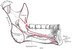
Mandible. Inner surface. Side view.
-

Hypoglossal nerve, cervical plexus, and their branches.
| Dissection images |
|---|
|
References
- ↑ Singh, Inderbir (2009). Essentials of anatomy (2nd ed.). New Delhi: Jaypee Bros. p. 348. ISBN 978-81-8448-461-8.
- 1 2 Drake, Richard L.; Vogl, Wayne; Tibbitts, Adam W.M. Mitchell (2005). Gray's anatomy for students. Philadelphia: Elsevier/Churchill Livingstone. pp. 991–2. ISBN 978-0-443-06612-2.
- ↑ Mu, Liancai; Sanders, Ira (2000). "Neuromuscular specializations of the pharyngeal dilator muscles: II. Compartmentalization of the canine genioglossus muscle". The Anatomical Record 260 (3): 308–25. doi:10.1002/1097-0185(20001101)260:3<308::aid-ar70>3.0.co;2-n. PMID 11066041.
- ↑ den Herder, Cindy; Schmeck, Joachim; Appelboom, Dick J K; de Vries, Nico (2004). "Risks of general anaesthesia in people with obstructive sleep apnoea". BMJ 329 (7472): 955–9. doi:10.1136/bmj.329.7472.955. PMC 524108. PMID 15499112.
External links
- Anatomy figure: 34:02-01 at Human Anatomy Online, SUNY Downstate Medical Center
- -227868593 at GPnotebook
- Anatomy diagram: 25420.000-1 at Roche Lexicon - illustrated navigator, Elsevier
- Frontal section
| ||||||||||||||||||||||||||||||||||||||||||||||||||||||||