p21
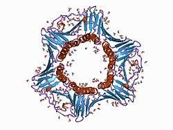
p21Cip1 (alternatively p21Waf1), also known as cyclin-dependent kinase inhibitor 1 or CDK-interacting protein 1, is a cyclin-dependent kinase inhibitor that inhibits the complexes of CDK2 and CDK1. This protein is encoded by the CDKN1A gene located on chromosome 6 (6p21.2) in humans.[1][2][3][4]
Function
p21 is a potent cyclin-dependent kinase inhibitor (CKI). The p21 (CIP1/WAF1) protein binds to and inhibits the activity of cyclin-CDK2, -CDK1, and -CDK4/6 complexes, and thus functions as a regulator of cell cycle progression at G1 and S phase.[5] In addition to growth arrest, p21 can mediate cellular senescence. One of the ways it was discovered was as a senescent cell-derived inhibitor.
The expression of this gene is tightly controlled by the tumor suppressor protein p53, through which this protein mediates the p53-dependent cell cycle G1 phase arrest in response to a variety of stress stimuli.[6] This was a major discovery in the early 1990s that revealed how cells stop dividing after being exposed to damaging agents such as radiation.
Studies of human embryonic stem cells (hESCs) commonly report the nonfunctional p53-p21 axis of the G1/S checkpoint pathway, and its relevance for cell cycle regulation and the DNA damage response (DDR). p21 mRNA is clearly present and upregulated after the DDR in hESCs, but p21 protein is not detectable. In this cell type, p53 activates numerous microRNAs (like miR-302a, miR-302b, miR-302c, and miR-302d) that directly inhibit the p21 expression in hESCs.[7]
p21 can also interact with proliferating cell nuclear antigen (PCNA), a DNA polymerase accessory factor, and plays a regulatory role in S phase DNA replication and DNA damage repair.[8] This protein was reported to be specifically cleaved by CASP3-like caspases, which thus leads to a dramatic activation of CDK2, and may be instrumental in the execution of apoptosis following caspase activation. However p21 may inhibit apoptosis and does not induce cell death on its own.[9] Two alternatively spliced variants, which encode an identical protein, have been reported.
Sometimes p21 is expressed without being induced by p53. This kind of induction plays a big role in p53 independent differentiation which is promoted by p21. Expression of p21 is mainly dependent on two factors 1) stimulus provided 2) type of the cell. Growth arrest by p21 can promote cellular differentiation. p21 therefore prevents cell proliferation.
Despite regulation by tumor suppressor gene p53, loss-of-function mutations in p21 (unlike p53) do not accumulate in cancer nor do they predispose to cancer incidence. Mice genetically engineered to lack p21 develop normally and are not susceptible to cancer at a higher rate than wild-type mice (unlike p53 knockout mice).
Mice that lack the p21 gene gain the ability to regenerate lost appendages.[10]
Clinical significance
p21 mediates the resistance of hematopoietic cells to an infection with HIV[11] by complexing with the HIV integrase and thereby aborting chromosomal integration of the provirus. HIV infected individuals who naturally suppress viral replication have elevated levels of p21 and its associated mRNA. p21 expression affects at least two stages in the HIV life cycle inside CD4 T cells, significantly limiting production of new viruses.[12]
Metastatic canine mammary tumors display increased levels of p21 in the primary tumors but also in their metastases, despite increased cell proliferation.[13][14]
Interactions
P21 has been shown to interact with:
References
- ↑ "Entrez Gene: CDKN1A cyclin-dependent kinase inhibitor 1A (p21, Cip1)".
- 1 2 Harper JW, Adami GR, Wei N, Keyomarsi K, Elledge SJ (November 1993). "The p21 Cdk-interacting protein Cip1 is a potent inhibitor of G1 cyclin-dependent kinases". Cell 75 (4): 805–16. doi:10.1016/0092-8674(93)90499-G. PMID 8242751.
- ↑ el-Deiry WS, Tokino T, Velculescu VE, Levy DB, Parsons R, Trent JM, Lin D, Mercer WE, Kinzler KW, Vogelstein B (November 1993). "WAF1, a potential mediator of p53 tumor suppression". Cell 75 (4): 817–25. doi:10.1016/0092-8674(93)90500-P. PMID 8242752.
- ↑ "p21 is a universal inhibitor of cyclin kinases.". Nature 366: 701–4. Dec 1993. doi:10.1038/366701a0. PMID 8259214.
- ↑ Gartel AL, Radhakrishnan SK (May 2005). "Lost in transcription: p21 repression, mechanisms, and consequences". Cancer Res. 65 (10): 3980–5. doi:10.1158/0008-5472.CAN-04-3995. PMID 15899785.
- ↑ Rodriguez R, Meuth M (January 2006). "Chk1 and p21 cooperate to prevent apoptosis during DNA replication fork stress". Mol. Biol. Cell 17 (1): 402–12. doi:10.1091/mbc.E05-07-0594. PMC 1345677. PMID 16280359.
- ↑ Dolezalova D, Mraz M, Barta T, Plevova K, Vinarsky V, Holubcova Z, Jaros J, Dvorak P, Pospisilova S, Hampl A (2012). "MicroRNAs regulate p21(Waf1/Cip1) protein expression and the DNA damage response in human embryonic stem cells". Stem Cells 30 (7): 1362–72. doi:10.1002/stem.1108. PMID 22511267.
- ↑ Xiong Y, Zhang H, Beach D (1992). "D type cyclins associate with multiple protein kinases and the DNA replication and repair factor PCNA". Cell 71 (3): 505–14. doi:10.1016/0092-8674(92)90518-h. PMID 1358458.
- ↑ Almond JB, Cohen GM (April 2002). "The proteasome: a novel target for cancer chemotherapy". Leukemia 16 (4): 433–43. doi:10.1038/sj.leu.2402417. PMID 11960320.
- ↑ Bedelbaeva K, Snyder A, Gourevitch D, Clark L, Zhang XM, Leferovich J, Cheverud JM, Lieberman P, Heber-Katz E (March 2010). "Lack of p21 expression links cell cycle control and appendage regeneration in mice". Proc. Natl. Acad. Sci. U.S.A. 107 (13): 5845–50. doi:10.1073/pnas.1000830107. PMC 2851923. PMID 20231440. Lay summary – PhysOrg.com.
- ↑ Zhang J, Scadden DT, Crumpacker CS (February 2007). "Primitive hematopoietic cells resist HIV-1 infection via p21". J. Clin. Invest. 117 (2): 473–81. doi:10.1172/JCI28971. PMC 1783820. PMID 17273559.
- ↑ Chen H, Li C, Huang J, Cung T, Seiss K, Beamon J, Carrington MF, Porter LC, Burke PS, Yang Y, Ryan BJ, Liu R, Weiss RH, Pereyra F, Cress WD, Brass AL, Rosenberg ES, Walker BD, Yu XG, Lichterfeld M (April 2011). "CD4+ T cells from elite controllers resist HIV-1 infection by selective upregulation of p21". J. Clin. Invest. 121 (4): 1549–60. doi:10.1172/JCI44539. PMID 21403397. Lay summary – Harvard Gazette.
- ↑ Klopfleisch R, Gruber AD (August 2009). "Differential expression of cell cycle regulators p21, p27 and p53 in metastasizing canine mammary adenocarcinomas versus normal mammary glands". Res. Vet. Sci. 87 (1): 91–6. doi:10.1016/j.rvsc.2008.12.010. PMID 19185891.
- ↑ Klopfleisch R, von Euler H, Sarli G, Pinho SS, Gärtner F, Gruber AD (2011). "Molecular carcinogenesis of canine mammary tumors: news from an old disease". Vet. Pathol. 48 (1): 98–116. doi:10.1177/0300985810390826. PMID 21149845.
- ↑ Chen W, Sun Z, Wang XJ, Jiang T, Huang Z, Fang D, Zhang DD (June 2009). "Direct interaction between Nrf2 and p21(Cip1/WAF1) upregulates the Nrf2-mediated antioxidant response". Mol. Cell 34 (6): 663–73. doi:10.1016/j.molcel.2009.04.029. PMC 2714804. PMID 19560419.
- 1 2 Ono T, Kitaura H, Ugai H, Murata T, Yokoyama KK, Iguchi-Ariga SM, Ariga H (October 2000). "TOK-1, a novel p21Cip1-binding protein that cooperatively enhances p21-dependent inhibitory activity toward CDK2 kinase". J. Biol. Chem. 275 (40): 31145–54. doi:10.1074/jbc.M003031200. PMID 10878006.
- ↑ Mitsui K, Matsumoto A, Ohtsuka S, Ohtsubo M, Yoshimura A (October 1999). "Cloning and characterization of a novel p21(Cip1/Waf1)-interacting zinc finger protein, ciz1". Biochem. Biophys. Res. Commun. 264 (2): 457–64. doi:10.1006/bbrc.1999.1516. PMID 10529385.
- 1 2 3 Abbas T, Sivaprasad U, Terai K, Amador V, Pagano M, Dutta A (September 2008). "PCNA-dependent regulation of p21 ubiquitylation and degradation via the CRL4Cdt2 ubiquitin ligase complex". Genes Dev. 22 (18): 2496–506. doi:10.1101/gad.1676108. PMC 2546691. PMID 18794347.
- 1 2 McKenzie PP, Danks MK, Kriwacki RW, Harris LC (July 2003). "P21Waf1/Cip1 dysfunction in neuroblastoma: a novel mechanism of attenuating G0-G1 cell cycle arrest". Cancer Res. 63 (13): 3840–4. PMID 12839982.
- ↑ Law BK, Chytil A, Dumont N, Hamilton EG, Waltner-Law ME, Aakre ME, Covington C, Moses HL (December 2002). "Rapamycin potentiates transforming growth factor beta-induced growth arrest in nontransformed, oncogene-transformed, and human cancer cells". Mol. Cell. Biol. 22 (23): 8184–98. doi:10.1128/MCB.22.23.8184-8198.2002. PMC 134072. PMID 12417722.
- ↑ Yam CH, Ng RW, Siu WY, Lau AW, Poon RY (January 1999). "Regulation of cyclin A-Cdk2 by SCF component Skp1 and F-box protein Skp2". Mol. Cell. Biol. 19 (1): 635–45. PMC 83921. PMID 9858587.
- ↑ Zhao H, Jin S, Antinore MJ, Lung FD, Fan F, Blanck P, Roller P, Fornace AJ, Zhan Q (July 2000). "The central region of Gadd45 is required for its interaction with p21/WAF1". Exp. Cell Res. 258 (1): 92–100. doi:10.1006/excr.2000.4906. PMID 10912791.
- ↑ Yang Q, Manicone A, Coursen JD, Linke SP, Nagashima M, Forgues M, Wang XW (November 2000). "Identification of a functional domain in a GADD45-mediated G2/M checkpoint". J. Biol. Chem. 275 (47): 36892–8. doi:10.1074/jbc.M005319200. PMID 10973963.
- ↑ Azam N, Vairapandi M, Zhang W, Hoffman B, Liebermann DA (January 2001). "Interaction of CR6 (GADD45gamma ) with proliferating cell nuclear antigen impedes negative growth control". J. Biol. Chem. 276 (4): 2766–74. doi:10.1074/jbc.M005626200. PMID 11022036.
- ↑ Nakayama K, Hara T, Hibi M, Hirano T, Miyajima A (August 1999). "A novel oncostatin M-inducible gene OIG37 forms a gene family with MyD118 and GADD45 and negatively regulates cell growth". J. Biol. Chem. 274 (35): 24766–72. doi:10.1074/jbc.274.35.24766. PMID 10455148.
- ↑ Rual JF, Venkatesan K, Hao T, Hirozane-Kishikawa T, Dricot A, Li N, Berriz GF, Gibbons FD, Dreze M, Ayivi-Guedehoussou N, Klitgord N, Simon C, Boxem M, Milstein S, Rosenberg J, Goldberg DS, Zhang LV, Wong SL, Franklin G, Li S, Albala JS, Lim J, Fraughton C, Llamosas E, Cevik S, Bex C, Lamesch P, Sikorski RS, Vandenhaute J, Zoghbi HY, Smolyar A, Bosak S, Sequerra R, Doucette-Stamm L, Cusick ME, Hill DE, Roth FP, Vidal M (October 2005). "Towards a proteome-scale map of the human protein-protein interaction network". Nature 437 (7062): 1173–8. doi:10.1038/nature04209. PMID 16189514.
- ↑ Frouin I, Maga G, Denegri M, Riva F, Savio M, Spadari S, Prosperi E, Scovassi AI (October 2003). "Human proliferating cell nuclear antigen, poly(ADP-ribose) polymerase-1, and p21waf1/cip1. A dynamic exchange of partners". J. Biol. Chem. 278 (41): 39265–8. doi:10.1074/jbc.C300098200. PMID 12930846.
- ↑ Watanabe H, Pan ZQ, Schreiber-Agus N, DePinho RA, Hurwitz J, Xiong Y (February 1998). "Suppression of cell transformation by the cyclin-dependent kinase inhibitor p57KIP2 requires binding to proliferating cell nuclear antigen". Proc. Natl. Acad. Sci. U.S.A. 95 (4): 1392–7. doi:10.1073/pnas.95.4.1392. PMC 19016. PMID 9465025.
- ↑ Fotedar R, Mossi R, Fitzgerald P, Rousselle T, Maga G, Brickner H, Messier H, Kasibhatla S, Hübscher U, Fotedar A (August 1996). "A conserved domain of the large subunit of replication factor C binds PCNA and acts like a dominant negative inhibitor of DNA replication in mammalian cells". EMBO J. 15 (16): 4423–33. PMC 452166. PMID 8861969.
- ↑ Jónsson ZO, Hindges R, Hübscher U (April 1998). "Regulation of DNA replication and repair proteins through interaction with the front side of proliferating cell nuclear antigen". EMBO J. 17 (8): 2412–25. doi:10.1093/emboj/17.8.2412. PMC 1170584. PMID 9545252.
- ↑ Gulbis JM, Kelman Z, Hurwitz J, O'Donnell M, Kuriyan J (October 1996). "Structure of the C-terminal region of p21(WAF1/CIP1) complexed with human PCNA". Cell 87 (2): 297–306. doi:10.1016/S0092-8674(00)81347-1. PMID 8861913.
- ↑ Touitou R, Richardson J, Bose S, Nakanishi M, Rivett J, Allday MJ (May 2001). "A degradation signal located in the C-terminus of p21WAF1/CIP1 is a binding site for the C8 alpha-subunit of the 20S proteasome". EMBO J. 20 (10): 2367–75. doi:10.1093/emboj/20.10.2367. PMC 125454. PMID 11350925.
- ↑ Yu P, Huang B, Shen M, Lau C, Chan E, Michel J, Xiong Y, Payan DG, Luo Y (January 2001). "p15(PAF), a novel PCNA associated factor with increased expression in tumor tissues". Oncogene 20 (4): 484–9. doi:10.1038/sj.onc.1204113. PMID 11313979.
- ↑ Wang Z, Bhattacharya N, Mixter PF, Wei W, Sedivy J, Magnuson NS (December 2002). "Phosphorylation of the cell cycle inhibitor p21Cip1/WAF1 by Pim-1 kinase". Biochim. Biophys. Acta 1593 (1): 45–55. doi:10.1016/S0167-4889(02)00347-6. PMID 12431783.
- ↑ Huang DY, Chang ZF (June 2001). "Interaction of human thymidine kinase 1 with p21(Waf1)". Biochem. J. 356 (Pt 3): 829–34. doi:10.1042/0264-6021:3560829. PMC 1221910. PMID 11389691.
- ↑ Oh H, Mammucari C, Nenci A, Cabodi S, Cohen SN, Dotto GP (April 2002). "Negative regulation of cell growth and differentiation by TSG101 through association with p21(Cip1/WAF1)". Proc. Natl. Acad. Sci. U.S.A. 99 (8): 5430–5. doi:10.1073/pnas.082123999. PMC 122786. PMID 11943869.
Further reading
- Marone M, Bonanno G, Rutella S, Leone G, Scambia G, Pierelli L (2002). "Survival and cell cycle control in early hematopoiesis: role of bcl-2, and the cyclin dependent kinase inhibitors P27 and P21". Leuk. Lymphoma 43 (1): 51–7. doi:10.1080/10428190210195. PMID 11908736.
- Fang JY, Lu YY (2002). "Effects of histone acetylation and DNA methylation on p21( WAF1) regulation". World J. Gastroenterol. 8 (3): 400–5. PMID 12046058.
- Tokumoto M, Tsuruya K, Fukuda K, Kanai H, Kuroki S, Hirakata H, Iida M (2003). "Parathyroid cell growth in patients with advanced secondary hyperparathyroidism: vitamin D receptor and cyclin-dependent kinase inhibitors, p21 and p27". Nephrol. Dial. Transplant. 18 Suppl 3: iii9–12. doi:10.1093/ndt/gfg1003. PMID 12771291.
- Amini S, Khalili K, Sawaya BE (2004). "Effect of HIV-1 Vpr on cell cycle regulators". DNA Cell Biol. 23 (4): 249–60. doi:10.1089/104454904773819833. PMID 15142382.
- Zhang Z, Wang H, Li M, Rayburn E, Agrawal S, Zhang R (2005). "Novel MDM2 p53-independent functions identified through RNA silencing technologies". Ann. N. Y. Acad. Sci. 1058: 205–14. doi:10.1196/annals.1359.030. PMID 16394138.
- P. Sankaranarayanan, T. E. Schomay, K. A. Aiello and O. Alter (April 2015). "Tensor GSVD of Patient- and Platform-Matched Tumor and Normal DNA Copy-Number Profiles Uncovers Chromosome Arm-Wide Patterns of Tumor-Exclusive Platform-Consistent Alterations Encoding for Cell Transformation and Predicting Ovarian Cancer Survival". PLOS ONE 10 (4): e0121396. doi:10.1371/journal.pone.0121396. PMID 25875127. AAAS EurekAlert! Press Release and NAE Podcast Feature.
External links
- Cyclin-Dependent Kinase Inhibitor p21 at the US National Library of Medicine Medical Subject Headings (MeSH)
- Drosophila dacapo - The Interactive Fly
- CDKN1A human gene location in the UCSC Genome Browser.
- CDKN1A human gene details in the UCSC Genome Browser.
| ||||||||||||||||||||||||||||||||||||||||||||||||||||||||||||||||||||||||||||||||||
| ||||||||||||||||||||||||||||||||||||||||

