Osteophyte
| Bone spur | |
|---|---|
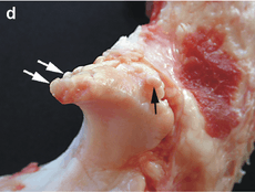
 Small marginal osteophytes (arrows) of the processus anconeus of the ulna can be seen in this gross pathological specimen of a sow. | |
| Classification and external resources | |
| Specialty | Orthopedics |
| ICD-10 | M25.7 |
| ICD-9-CM | 726.91 |
| DiseasesDB | 18621 |
| MeSH | D054850 |
Osteophytes, commonly referred to as bone spurs or parrot beak,[1] are bony projections that form along joint margins.[2] They should not be confused with enthesophytes, which are bony projections that form at the attachment of a tendon or ligament.[3]
Cause

Osteophyte formation has been classically related to any sequential and consequential changes in bone formation that is due to aging, degeneration, mechanical instability, and disease (such as Diffuse Idiopathic Skeletal Hyperostosis). Often osteophytes form in osteoarthritic joints as a result of damage and wear from inflammation. Calcification and new bone formation can also occur in response to mechanical damage in joints.[4]
Pathophysiology
Osteophytes form because of the increase in a damaged joint's surface area. This is most common from the onset of arthritis. Osteophytes usually limit joint movement and typically cause pain.[1]
Osteophytes form naturally on the back of the spine as a person ages and are a sign of degeneration in the spine. In this case, the spurs are not the source of back pains, but instead are the common symptom of a deeper problem. However, bone spurs on the spine can impinge on nerves that leave the spine for other parts of the body. This impingement can cause pain in both upper and lower limbs and a numbness or tingling sensations in the hands and feet because the nerves are supplying sensation to their dermatomes.[5]
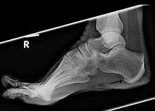
Spurs can also appear on the feet, either along toes or the heel, as well as on the hands. In extreme cases, bone spurs have grown along a person's entire skeletal structure: along the knees, hips, shoulders, ribs, arms and ankles. Such cases are only exhibited with multiple exostoses.
Osteophytes on the fingers or toes are known as Heberden's nodes (if on the distal interphalangeal joint) or Bouchard's nodes (if on the proximal interphalangeal joints).
Osteophytes may also be the end result of certain disease processes. Osteomyelitis, a bone infection, may leave the adjacent bone with a spur formation. Charcot foot, the neuropathic breakdown of the feet seen primarily in diabetics, can also leave bone spurs that may then become symptomatic.
Treatments
Normally, asymptomatic cases are not treated. Non-steroidal anti inflammatory drugs and surgery are two typical options for the rest.
Fossil record
Evidence for bone spurs found in the fossil record is studied by paleopathologists, specialists in ancient disease and injury. Bone spurs have been reported in dinosaur fossils from several species, including Allosaurus fragilis, Neovenator salerii, and Tyrannosaurus rex.[6]
References
- 1 2 Bone spurs MayoClinic.com
- ↑ "osteophyte" at Dorland's Medical Dictionary
- ↑ Rogers J, Shepstone L, Dieppe P (Feb 1997). "Bone formers: osteophyte and enthesophyte formation are positively associated". Annals of the Rheumatic Diseases 56 (2): 85–90. doi:10.1136/ard.56.2.85. PMC 1752321. PMID 9068279.
- ↑ Nathan M, Pope MH, Grobler LJ (Aug 1994). "Osteophyte formation in the vertebral column: a review of the etiologic factors--Part II". Contemporary Orthopaedics 29 (2): 113–9. PMID 10150240.
- ↑ Laser Spine Institute
- ↑ Molnar, R. E. (2001). "Theropod Paleopathology: A Literature Survey". In Tanke, D. H.; Carpenter, K. Mesozoic Vertebrate Life. Bloomington: Indiana University Press. pp. 337–363. ISBN 0-253-33907-3.
External links
- Mayo Clinic website concise information on bone spurs
- Laser Spine Institute information on spinal bone spurs or ostephytes
| ||||||||||||||||||||||
| ||||||||||||||||||||||||||||||||||||||||||