Auditory system

The auditory system is the sensory system for the sense of hearing. It includes both the sensory organs (the ears) and the auditory parts of the sensory system.
Peripheral auditory system
The auditory periphery, starting with the ear, is the first stage of the transduction of sound in a hearing organism. While not part of the nervous system, its components feed directly into the nervous system, performing mechanoeletrical transduction of sound pressure waves into neural action potentials.
Outer ear
The folds of cartilage surrounding the ear canal are called the pinna. Sound waves are reflected and attenuated when they hit the pinna, and these changes provide additional information that will help the brain determine the direction from which the sounds came.
The sound waves enter the auditory canal, a deceptively simple tube. The ear canal amplifies sounds that are between 3 and 12 kHz. At the far end of the ear canal is the tympanic membrane, which marks the beginning of the middle ear.
Middle ear
Sound waves travel through the ear canal and hit the tympanic membrane, or eardrum. This wave information travels across the air-filled middle ear cavity via a series of delicate bones: the malleus (hammer), incus (anvil) and stapes (stirrup). These ossicles act as a lever, converting the lower-pressure eardrum sound vibrations into higher-pressure sound vibrations at another, smaller membrane called the oval (or elliptical) window. The manubrium (handle) of the malleus articulates with the tympanic membrane, while the footplate of the stapes articulates with the oval window. Higher pressure is necessary at the oval window than at the typanic membrane because the inner ear beyond the oval window contains liquid rather than air. The stapedius reflex of the middle ear muscles helps protect the inner ear from damage by reducing the transmission of sound energy when the stapedius muscle is activated in response to sound. The middle ear still contains the sound information in wave form; it is converted to nerve impulses in the cochlea.
Inner ear
| Cochlea | |
|---|---|
|
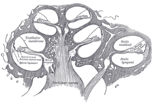
Diagrammatic longitudinal section of the cochlea. Scala media is labeled as ductus cochlearis at right. | |
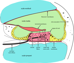
The inner ear consists of the cochlea and several non-auditory structures. The cochlea has three fluid-filled sections, and supports a fluid wave driven by pressure across the basilar membrane separating two of the sections. Strikingly, one section, called the cochlear duct or scala media, contains endolymph, a fluid similar in composition to the intracellular fluid found inside cells. The organ of Corti is located in this duct on the basilar membrane, and transforms mechanical waves to electric signals in neurons. The other two sections are known as the scala tympani and the scala vestibuli; these are located within the bony labyrinth, which is filled with fluid called perilymph, similar in composition to cerebrospinal fluid. The chemical difference between the fluids endolymph and perilymph fluids is important for the function of the inner ear due to electrical potential differences between potassium and calcium ions.
The plan view of the human cochlea (typical of all mammalian and most vertebrates) shows where specific frequencies occur along its length. The frequency is an approximately exponential function of the length of the cochlea within the Organ of Corti. In some species, such as bats and dolphins, the relationship is expanded in specific areas to support their active sonar capability.
Organ of Corti

The organ of Corti forms a ribbon of sensory epithelium which runs lengthwise down the cochlea's entire scala media. Its hair cells transform the fluid waves into nerve signals. The journey of countless nerves begins with this first step; from here, further processing leads to a panoply of auditory reactions and sensations.
Hair cell
Hair cells are columnar cells, each with a bundle of 100-200 specialized cilia at the top, for which they are named. There are two types of hair cells. Inner hair cells are the mechanoreceptors for hearing: they transduce the vibration of sound into electrical activity in nerve fibers, which is transmitted to the brain. Outer hair cells are a motor structure. Sound energy causes changes in the shape of these cells, which serves to amplify sound vibrations in a frequency specific manner. Lightly resting atop the longest cilia of the inner hair cells is the tectorial membrane, which moves back and forth with each cycle of sound, tilting the cilia, which is what elicits the hair cells' electrical responses.
Inner hair cells, like the photoreceptor cells of the eye, show a graded response, instead of the spikes typical of other neurons. These graded potentials are not bound by the “all or none” properties of an action potential.
At this point, one may ask how such a wiggle of a hair bundle triggers a difference in membrane potential. The current model is that cilia are attached to one another by “tip links”, structures which link the tips of one cilium to another. Stretching and compressing, the tip links may open an ion channel and produce the receptor potential in the hair cell. Recently it has been shown that Cadherin-23 CDH23 and Protocadherin-15 PCDH15 are the adhesion molecules associated with these tip links.[1] It is thought that a calcium driven motor causes a shortening of these links to regenerate tensions. This regeneration of tension allows for apprehension of prolonged auditory stimulation.[2]
Neurons
Afferent neurons innervate cochlear inner hair cells, at synapses where the neurotransmitter glutamate communicates signals from the hair cells to the dendrites of the primary auditory neurons.
There are far fewer inner hair cells in the cochlea than afferent nerve fibers – many auditory nerve fibers innervate each hair cell. The neural dendrites belong to neurons of the auditory nerve, which in turn joins the vestibular nerve to form the vestibulocochlear nerve, or cranial nerve number VIII.[3] The region of the basilar membrane supplying the inputs to a particular afferent nerve fibre can be considered to be its receptive field.
Efferent projections from the brain to the cochlea also play a role in the perception of sound, although this is not well understood. Efferent synapses occur on outer hair cells and on afferent (towards the brain) dendrites under inner hair cells
Central auditory system

This sound information, now re-encoded, travels down the vestibulocochlear nerve, through intermediate stations such as the cochlear nuclei and superior olivary complex of the brainstem and the inferior colliculus of the midbrain, being further processed at each waypoint. The information eventually reaches the thalamus, and from there it is relayed to the cortex. In the human brain, the primary auditory cortex is located in the temporal lobe.
Associated anatomical structures include:
Cochlear nucleus
The cochlear nucleus is the first site of the neuronal processing of the newly converted “digital” data from the inner ear (see also binaural fusion). In mammals, this region is anatomically and physiologically split into two regions, the dorsal cochlear nucleus (DCN), and ventral cochlear nucleus (VCN).
Trapezoid body
The Trapezoid body is a bundle of decussating fibers in the ventral pons that carry information used for binaural computations in the brainstem.
Superior olivary complex
The superior olivary complex is located in the pons, and receives projections predominantly from the ventral cochlear nucleus, although the dorsal cochlear nucleus projects there as well, via the ventral acoustic stria. Within the superior olivary complex lies the lateral superior olive (LSO) and the medial superior olive (MSO). The former is important in detecting interaural level differences while the latter is important in distinguishing interaural time difference.[4]
Lateral lemniscus
The lateral lemniscus is a tract of axons in the brainstem that carries information about sound from the cochlear nucleus to various brainstem nuclei and ultimately the contralateral inferior colliculus of the midbrain.
Inferior colliculi
The inferior colliculus (IC) are located just below the visual processing centers known as the superior colliculi. The central nucleus of the IC is a nearly obligatory relay in the ascending auditory system, and most likely acts to integrate information (specifically regarding sound source localization from the superior olivary complex[5] and dorsal cochlear nucleus) before sending it to the thalamus and cortex.[6]
Medial geniculate nucleus
The medial geniculate nucleus is part of the thalamic relay system.
Primary auditory cortex
The primary auditory cortex is the first region of cerebral cortex to receive auditory input.
Perception of sound is associated with the left posterior superior temporal gyrus (STG). The superior temporal gyrus contains several important structures of the brain, including Brodmann areas 41 and 42, marking the location of the primary auditory cortex, the cortical region responsible for the sensation of basic characteristics of sound such as pitch and rhythm. We know from work in nonhuman primates that primary auditory cortex can probably itself be divided further into functionally differentiable subregions.[7][8][9][10] [11][12][13] The neurons of the primary auditory cortex can be considered to have receptive fields covering a range of auditory frequencies and have selective responses to harmonic pitches.[14] Neurons integrating information from the two ears have receptive fields covering a particular region of auditory space.
Primary auditory cortex is surrounded by secondary auditory cortex, and interconnects with it. These secondary areas interconnect with further processing areas in the superior temporal gyrus, in the dorsal bank of the superior temporal sulcus, and in the frontal lobe. In humans, connections of these regions with the middle temporal gyrus are probably important for speech perception. The frontotemporal system underlying auditory perception allows us to distinguish sounds as speech, music, or noise.
See also
- Auditory brainstem response and ABR audiometry test for newborn hearing
- Auditory processing disorder
- Cognitive neuroscience of music
- Endaural phenomena
- Gammatone filter – a simple linear model of peripheral auditory filtering
- Noise health effects
- Selective auditory attention
- Sound
- Tinnitus
References
- ↑ Lelli, A.; Kazmierczak, P.; Kawashima, Y.; Muller, U.; Holt, J. R. (2010). "Development and Regeneration of Sensory Transduction in Auditory Hair Cells Requires Functional Interaction Between Cadherin-23 and Protocadherin-15". Journal of Neuroscience 30 (34): 11259–11269. doi:10.1523/JNEUROSCI.1949-10.2010. PMC 2949085. PMID 20739546.
- ↑ Peng, AW.; Salles, FT.; Pan, B.; Ricci, AJ. (2011). "Integrating the biophysical and molecular mechanisms of auditory hair cell mechanotransduction.". Nature Communications 2: 523–. doi:10.1038/ncomms1533. PMC 3418221. PMID 22045002.
- ↑ Meddean – CN VIII. Vestibulocochlear Nerve
- ↑ Moore JK (November 2000). <403::AID-JEMT8>3.0.CO;2-Q "Organization of the human superior olivary complex". Microsc. Res. Tech. 51 (4): 403–12. doi:10.1002/1097-0029(20001115)51:4<403::AID-JEMT8>3.0.CO;2-Q. PMID 11071722.
- ↑ Oliver DL (November 2000). <355::AID-JEMT5>3.0.CO;2-J "Ascending efferent projections of the superior olivary complex". Microsc. Res. Tech. 51 (4): 355–63. doi:10.1002/1097-0029(20001115)51:4<355::AID-JEMT5>3.0.CO;2-J. PMID 11071719.
- ↑ Demanez JP, Demanez L (2003). "Anatomophysiology of the central auditory nervous system: basic concepts". Acta Otorhinolaryngol Belg 57 (4): 227–36. PMID 14714940.
- ↑ Pandya DN (1995). "Anatomy of the auditory cortex". Rev. Neurol. (Paris) 151 (8-9): 486–94. PMID 8578069.
- ↑ Kaas JH, Hackett TA (1998). "Subdivisions of auditory cortex and levels of processing in primates". Audiol. Neurootol. 3 (2-3): 73–85. doi:10.1159/000013783. PMID 9575378.
- ↑ Kaas JH, Hackett TA, Tramo MJ (April 1999). "Auditory processing in primate cerebral cortex". Curr. Opin. Neurobiol. 9 (2): 164–70. doi:10.1016/S0959-4388(99)80022-1. PMID 10322185.
- ↑ Kaas JH, Hackett TA (October 2000). "Subdivisions of auditory cortex and processing streams in primates". Proc. Natl. Acad. Sci. U.S.A. 97 (22): 11793–9. doi:10.1073/pnas.97.22.11793. PMC 34351. PMID 11050211.
- ↑ Hackett TA, Preuss TM, Kaas JH (December 2001). "Architectonic identification of the core region in auditory cortex of macaques, chimpanzees, and humans". J. Comp. Neurol. 441 (3): 197–222. doi:10.1002/cne.1407. PMID 11745645.
- ↑ Scott SK, Johnsrude IS (February 2003). "The neuroanatomical and functional organization of speech perception". Trends Neurosci. 26 (2): 100–7. doi:10.1016/S0166-2236(02)00037-1. PMID 12536133.
- ↑ Tian B, Reser D, Durham A, Kustov A, Rauschecker JP (April 2001). "Functional specialization in rhesus monkey auditory cortex". Science 292 (5515): 290–3. doi:10.1126/science.1058911. PMID 11303104.
- ↑ Wang X (2013). "The harmonic organization of auditory cortex". Front Syst Neurosci 7: 114. doi:10.3389/fnsys.2013.00114. PMC 3865599. PMID 24381544.
Further reading
- Kandel, Eric R. (2012). Principles of Neural Science. New York: McGraw-Hill. ISBN 978-0-07-139011-8. OCLC 795553723.
External links
- Promenade 'round the cochlea
- Auditory system – Washington University Neuroscience Tutorial
- Lincoln Gray. "Chapter 13: Auditory System: Pathways and Reflexes". Neuroscience Online, the Open-Access Neuroscience Electronic Textbook. The University of Texas Health Science Center at Houston (UTHealth). Retrieved 27 April 2014.
| ||||||||||||||
| ||||||||||||||||||||||||||||||||||||||||||||||||||
| ||||||||||||||||||||||
| ||||||||||||||||||||||||||||||
