Third ventricle
| Third ventricle | |
|---|---|
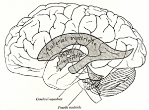 Scheme showing relations of the ventricles to the surface of the brain. | |
| Details | |
| Latin | ventriculus tertius cerebri |
| Identifiers | |
| Gray's | p.813 |
| NeuroNames | hier-429 |
| NeuroLex ID | Third ventricle |
| Dorlands /Elsevier | v_06/12853453 |
| TA | A14.1.08.410 |
| FMA | 78454 |
| Anatomical terms of neuroanatomy | |
The third ventricle (ventriculus tertius) is one of four connected fluid-filled cavities comprising the ventricular system within the human brain. It is a median cleft in the diencephalon between the two thalami, and is filled with cerebrospinal fluid (CSF).
It is in the midline, between the left and right lateral ventricles. Running through the third ventricle is the Interthalamic adhesion, which contains thalamic neurons and fibers that may connect the two thalami.
Structure
The third ventricle communicates with the lateral ventricles anteriorly by the interventricular foramina (of Monro). It also communicates with the fourth ventricle posteriorly by the cerebral aqueduct (of Sylvius).
Development
The third ventricle, like other parts of the ventricular system of the brain, develops from the central canal of the neural tube. Specifically, it originates from the portion of the tube that is present in the developing prosencephalon, and subsequently in the developing diencephalon.[1]
Boundaries

The third ventricle is bounded by the thalamus and hypothalamus on both the left and right sides. The lamina terminalis forms the anterior wall. The floor is formed by hypothalamic structures, and can be opened surgically between the mamillary bodies and the pituitary gland in a procedure called an endoscopic third ventriculostomy. The roof is formed by the ependyma, lining the undersurface of the tela choroidea of the third ventricle.
Protrusions
There are two protrusions on the anterior aspect of the third ventricle:
- the supra-optic recess (above the optic chiasma)
- the infundibular recess (above the pituitary stalk).
Additionally, there are two protrusions on the posterior aspect, above the cerebral aqueduct:
- the suprapineal recess (above the pineal gland)
- the pineal recess (protruding into the stalk of the pineal gland)
In casts of the ventricular system, a small hole may be seen in the body of the third ventricle. This is formed where the two thalami are joined together at the interthalamic adhesion (not seen in all people).
See also
- Biology of depression
- Suprapineal recess
- Tanycytes line the bottom of the ventricle
References
Additional images
-
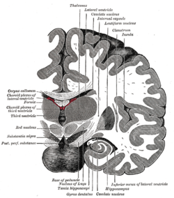
Coronal section of brain immediately in front of pons.
-
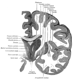
Coronal section of brain through intermediate mass of third ventricle.
-
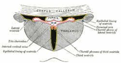
Coronal section of lateral and third ventricles.
-
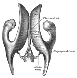
Drawing of a cast of the ventricular cavities, viewed from above.
-
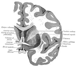
Coronal section of brain through anterior commissure.
-
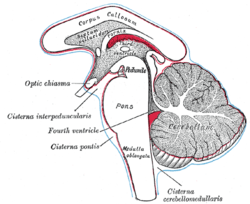
Diagram showing the positions of the three principal subarachnoid cisternæ.
-
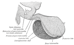
Median sagittal through the hypophysis of an adult monkey. Semidiagrammatic.
-
Third ventricle
-
Third ventricle
External links
| Wikimedia Commons has media related to Third ventricle. |
| ||||||||||||||||||||||||||||

