Temporal horn of lateral ventricle
| Temporal horn of lateral ventricle | |
|---|---|
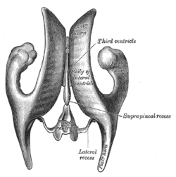 Drawing of a cast of the ventricular cavities, viewed from above. | |
 Drawing of a cast of the ventricular cavities, viewed from the side. | |
| Details | |
| Latin | cornu inferior |
| Identifiers | |
| Gray's | p.829 |
| NeuroNames | hier-204 |
| NeuroLex ID | Inferior horn of the lateral ventricle |
| TA | A14.1.09.287 |
| FMA | 83701 |
| Anatomical terms of neuroanatomy | |
The temporal horn of lateral ventricle (inferior horn, descending horn; middle horn; medicornu), the largest of the horns of the lateral ventricle, traverses the temporal lobe of the brain, forming in its course a curve around the posterior end of the thalamus.
It passes at first backward, lateralward, and downward, and then curves forward to within 2.5 cm. of the apex of the temporal lobe, its direction being fairly well indicated on the surface of the brain by that of the superior temporal sulcus.
Its roof is formed chiefly by the inferior surface of the tapetum of the corpus callosum, but the tail of the caudate nucleus and the stria terminalis also extend forward in the roof of the temporal horn to its extremity; the tail of the caudate nucleus joins the putamen.
Its floor presents the following parts: the hippocampus, the fimbria hippocampi, the collateral eminence, and the choroid plexus.
When the choroid plexus is removed, a cleft-like opening is left along the medial wall of the temporal horn; this cleft constitutes the lower part of the choroidal fissure.
Additional images
-
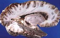
Human brain right dissected lateral view
References
This article incorporates text in the public domain from the 20th edition of Gray's Anatomy (1918)
External links
- Atlas image: n1a4p2 at the University of Michigan Health System
- Atlas image: n1a4p5 at the University of Michigan Health System
| ||||||||||||||||||||||||||||