T7 phage
| Enterobacteria phage T7 | |
|---|---|
| Virus classification | |
| Group: | Group I (dsDNA) |
| Order: | Caudovirales |
| Family: | Podoviridae |
| Genus: | T7-like viruses |
| Species: | T7 phage |
Bacteriophage T7 is a bacteriophage capable of infecting susceptible bacterial cells. It infects most strains of Escherichia coli (including E. coli O157:H7, (this needs a citation) - a strain of E. coli which can cause foodborne illness). Bacteriophage T7 is a DNA virus that has a lytic life cycle. The T7 bacteriophage has several properties that make it an ideal phage for experimentation.
Discovery
Bacteriophage T7 was identified in 1945 as a member of the seven Type (“T”) phages that grow lytically on Escherichia coli B,[1] although it is probably identical to phage δ, used earlier by Delbrück. A close relative of T7 was likely studied by d’Herelle in the 1920s.[2]
Hosts
T7 grows on rough strains of E. coli (i.e. those without full-length O-antigen polysaccharide on their surface) and some other enteric bacteria, but close relatives also infect smooth and even capsulated strains.[3]
Virion structure
The virus has complex structural symmetry, with a capsid of the phage that is icosahedral with an inner diameter of 55 nm and a tail 19 nm in diameter and 28.5 nm long attached to the capsid.
Genome
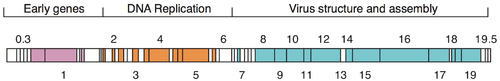
The genome of phage T7 [4] was among the first completely sequenced genomes and was published in 1983.[5] The head of the phage particle contains the roughly 40 kbp dsDNA genome which encodes 55 proteins.[6]
Life cycle
T7 has a short life cycle of 17 min at 37˚C, i.e. the time from infection to the lysis of the host cell when new phage are released. Due to the short latent period most physiological studies are being conducted at 30˚C where infected cells lyse after 30 min. However, high fitness-strains of T7 have been isolated with a latent period of only ~11 min at 37˚C growing under optimal conditions in rich media results. This adapted phage can undergo an effective expansion of its population by more than 1013 in one hour of growth.[7]
Infection of host bacteria
The T7 phage recognizes receptors on the surface of E.coli cells. It adheres to the cell surface by the tail fibers. In some strains of T7 the tail fibers are replaced with tailspikes with enzymatic activity that degrades the O- or K-antigens on the cell surface. The adsorbtion and penetration process use such lysozymes to create an opening within the peptidoglycan layer of the bacterial cell wall allowing transfer of the viral DNA into the bacterium. The short, stubby tail of the T7-like phages is too short to span the cell envelope and, in order to eject the phage genome into the cell at the initiation of infection, virion proteins must first make a channel from the tip of the tail into the cell cytoplasm.[8] The phage also injects proteins needed to begin replication of the viral genome and cleave the host genome.[9] T7 bacteriophage overcomes several of the host bacteria's defenses including the peptidoglycan cell wall and the CRISPR system.[9] Once the T7 phage has inserted the viral genome the process of DNA replication of the host genome is halted and replication of viral genome begins. The T7 phage completes the lytic process in optimal conditions within 25 minutes. At the time of lysis the virus can produce over 100 progeny.[9]
Components
Gp5 (encoded by gene gp5) is T7 phage's DNA polymerase. T7 polymerase uses E. coli's endogenous thioredoxin as a sliding clamp during phage DNA replication (though thioredoxin normally has a different function). The sliding clamp functions to hold the polymerase onto the DNA, which increases the rate of synthesis; initiation, the process by which a polymerase binds to DNA, is time-consuming.
Applications in molecular biology
The T7 promoter sequence is used extensively in molecular biology due to its extremely high affinity for T7 RNA polymerase and thus high level of expression.
T7 has been used as a model in synthetic biology. Chan et al. (2005) "refactored" the genome of T7, replacing approximately 12 kbp of its genome with engineered DNA.[10] The engineered DNA was designed to be easier to work with in a number of ways: individual functional elements were separated by restriction endonuclease sites for simple modification, and overlapping protein coding domains were separated and, where necessary, modified by single base pair silent mutations. T7 has been tested on human osteosarcoma to treat tumor cells. Chen et al. (1998)"To test the utility of the system in vivo tumor ablation, a T7 cancer gene therapy plasmid vector, pT7T7/T7TK, was constructed. This nonviral vector contains a T7 autogene, T7T7, and a human herpes simplex virus thymidine kinase (HSV-TK) gene driven by a second T7 promoter (T7TK). When co-transfected with T7 RNA polymerase (T7 RNAP) into cultured human osteosarcoma 143B cells, about 10-20% of the cells were found to express HSV-TK, and more than 90% of the cells were killed in the presence of 1 microM ganciclovir (GCV) within 4 days after DNA transfection."[11]
References
- ↑ Demerec, M.; Fano, U. (1945). "Bacteriophage-Resistant Mutants in Escherichia Coli". Genetics 30 (2): 119–136. PMC 1209279. PMID 17247150.
- ↑ d’Herelle, F. (1926). The Bacteriophage and Its Behavior. Baltimore, MD: Williams & Wilkins
- ↑ Molineux, I. J. (2006). Chapter 20: The T7 group. In: The Bacteriophages (R. Calendar, ed.), pp. 277. Oxford University Press, Oxford.
- ↑ "Genome of bacteriophage T7".
- ↑ "Dunn,J.J. and Studier,F.W. (1983) Complete nucleotide sequence of bacteriophage T7 DNA and the locations of T7 genetic elements. J. Mol. Biol. 166 (4), 477-535".
- ↑ "Uniprot: reference proteome of bacteriophage T7".
- ↑ Heineman, R. H.; Bull, J. J. (2007). "Testing Optimality with Experimental Evolution: Lysis Time in a Bacteriophage". Evolution 61 (7): 1695–1709. doi:10.1111/j.1558-5646.2007.00132.x. PMC 1974807. PMID 17598749.
- ↑ "Gp15 and gp16 cooperate in translocating bacteriophage T7 DNA into the infected cell". Virology 398 (2). 2010. pp. 176–186.
- ↑ 9.0 9.1 9.2 "New Details about Bacteriophage T7-Host Interactions".
- ↑ Leon Y. Chan, Sriram Kosuri, Drew Endy (2005). "Refactoring bacteriophage T7". Molecular Systems Biology 1 (1): 2005.0018. doi:10.1038/msb4100025. PMC 1681472. PMID 16729053.
- ↑ Xiaozhuo Chen, Li, Xiong,Aizicovici, Xie,Zhu,Sturtz, Shulok,Snodgrass, Wagner,Platika, (March 2008). "Cancer Gene Therapy by Direct Tumor Injections of a Nonviral T7 Vector Encoding a Thymidine Kinase Gene". Human Gene Therapy 9 (5): 729–736. doi:10.1089/hum.1998.9.5-729. PMID 9551620. Retrieved 2012.
External links
- T7 Phage at the US National Library of Medicine Medical Subject Headings (MeSH)
- ViralZone
- New Details about T7-host interactions. Microbe Magazine
- T7 reference proteome in Uniprot