Superior ganglion of vagus nerve
| Superior ganglion of vagus nerve | |
|---|---|
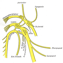 Plan of upper portions of glossopharyngeal, vagus, and accessory nerves. (Jugular ganglion visible near center.) | |
| Details | |
| Latin | ganglion superius nervi vagi, ganglion jugulare |
| Identifiers | |
| Gray's | p.911 |
| Dorlands /Elsevier | g_02/12385029 |
| TA | A14.2.01.154 |
| Anatomical terms of neuroanatomy | |
The vagus presents a well-marked ganglionic enlargement, which is called the superior ganglion of the vagus nerve (also known as jugular ganglion as it sits in the jugular foramen). It contains afferent somatosensory neuronal cell bodies that provide sensory information from the external auditory meatus (auricular branch), cranial meninges (meningeal branch), and the external surface of the tympanic membrane. Their central fibers synapse in the sensory trigeminal nucleus.[1]
It is a grayish color, spherical in form, and about 4 mm. in diameter.
References
This article incorporates text in the public domain from the 20th edition of Gray's Anatomy (1918)
- ↑ Netter's Human Anatomy, 4th Edition
See also
External links
- cranialnerves at The Anatomy Lesson by Wesley Norman (Georgetown University) (X)
- ‹The template EMedicineDictionary is being considered for deletion.› superior+ganglion+of+vagus+nerve at eMedicine Dictionary
| ||||||||||||||||||||||||||||||||||||||||||||||||||||||||||||||||||||||||||||||||||||||||||||||||||||||||||||||||||||||