Stomach
| Stomach | |
|---|---|
 The stomach is located centre left in the human body. | |
 1. Body of stomach 2. Fundus 3. Anterior wall 4. Greater curvature 5. Lesser curvature 6. Cardia 9. Pyloric sphincter 10. Pyloric antrum 11. Pyloric canal 12. Angular incisure 13. Gastric canal 14. Rugae[1] | |
| Details | |
| Latin | Ventriculus |
| Greek | Gaster |
| Precursor | Foregut |
| System | Digestive system |
| Right gastric artery, left gastric artery, right gastro-omental artery, left gastro-omental artery, short gastric arteries | |
| Right gastric vein, left gastric vein, right gastroepiploic vein, left gastroepiploic vein, short gastric veins | |
| Celiac ganglia, vagus nerve[2] | |
| Celiac lymph nodes[3] | |
| Identifiers | |
| Gray's | p.1161 |
| MeSH | A03.556.875.875 |
| Dorlands /Elsevier | Stomach |
| TA | A05.5.01.001 |
| FMA | 7148 |
| Anatomical terminology | |
The stomach is a muscular, hollow, dilated part of the digestive system which functions as an important organ of the digestive tract in some animals, including vertebrates, echinoderms, insects (mid-gut), and molluscs. It is involved in the second phase of digestion, following mastication (chewing).
In most vertebrates, the stomach is located between the esophagus and the small intestine. It secretes protein-digesting enzymes called proteases and strong acids to aid in food digestion, (sent to it via esophageal peristalsis) through smooth muscular contortions (called segmentation) before sending partially digested food (chyme) to the small intestines.
Human stomach
Structure

The stomach lies between the esophagus and the duodenum (the first part of the small intestine). It is on the left upper part of the abdominal cavity. The top of the stomach lies against the diaphragm. Lying behind the stomach is the pancreas. The greater omentum hangs down from the greater curvature.
Two sphincters keep the contents of the stomach contained. They are the lower esophageal sphincter or Cardiac sphincter (found in the cardiac region, not an anatomical sphincter) dividing the tract above, and the pyloric sphincter dividing the stomach from the small intestine.
The stomach is surrounded by parasympathetic (stimulant) and orthosympathetic (inhibitor) plexuses (networks of blood vessels and nerves in the anterior gastric, posterior, superior and inferior, celiac and myenteric), which regulate both the secretions activity and the motor (motion) activity of its muscles.
In adult humans, the stomach has a relaxed, near empty volume of about 45 to 75 milliliters.[4] Because it is a distensible organ, it normally expands to hold about one litre of food.[5] The stomach of a newborn human baby will only be able to retain about 30 milliliters.
Sections
In classical anatomy, the stomach is divided into four sections, beginning at the cardia,[6] each of which has different cells and functions.
- The cardia is where the contents of the esophagus empty into the stomach. The cardia is defined as the region following the "z-line" of the gastroesophageal junction, the point at which the epithelium changes from stratified squamous to columnar. Near the cardia is the lower esophageal sphincter.[7]
- The fundus is formed by the upper curvature of the organ.
- The body (Latin: corpus) is the main, central region.
- The Pylorus is the lower section of the organ that facilitates emptying the contents into the small intestine.
Blood supply
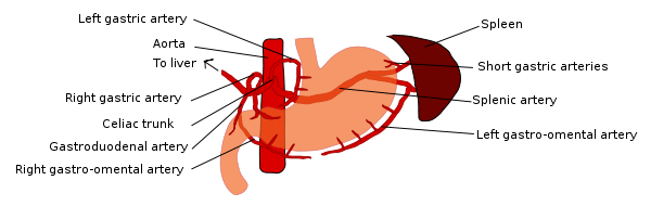
The lesser curvature of the stomach is supplied by the right gastric artery inferiorly, and the left gastric artery superiorly, which also supplies the cardiac region. The greater curvature is supplied by the right gastro-omental artery inferiorly and the left gastro-omental artery superiorly. The fundus of the stomach, and also the upper portion of the greater curvature, is supplied by the short gastric artery which arises from the splenic artery.
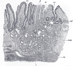
Histology

Like the other parts of the gastrointestinal tract, the stomach walls consist of an outer mucosa, and inner submucosa, muscularis externa, and serosa.
The mucosa of the stomach consists of the epithelium and the lamina propria (composed of loose connective tissue), with a thin layer of smooth muscle called the muscularis mucosae separating it from the submucosa beneath. The submucosa lies under the mucosa and consists of fibrous connective tissue, separating the mucosa from the next layer. Meissner's plexus is in this layer. The muscularis externa lies beneath the submucosa, and is unique from other organs of the gastrointestinal tract, consisting of three layers:
- The inner oblique layer: This layer is responsible for creating the motion that churns and physically breaks down the food. It is the only layer of the three which is not seen in other parts of the digestive system. The antrum has thicker skin cells in its walls and performs more forceful contractions than the fundus.
- The middle circular layer: At this layer, the pylorus is surrounded by a thick circular muscular wall which is normally tonically constricted forming a functional (if not anatomically discrete) pyloric sphincter, which controls the movement of chyme into the duodenum. This layer is concentric to the longitudinal axis of the stomach.
- Auerbach's plexus (AKA myenteric plexus) is found between the outer longitundinal and the middle circular layer and is responsible for the innervation of both (causing peristalsis and mixing)
- The outer longitudinal layer
The stomach also possesses a serosa, consisting of layers of connective tissue continuous with the peritoneum.
Glands
Different types of cells are found at the different layers of these glands:
| Layer of stomach | Name | Secretion | Region of stomach | Staining |
| Isthmus of gland | Foveolar cells | mucus gel layer | Fundic, cardiac, pyloric | Clear |
| Body of gland | parietal (oxyntic) cells | gastric acid and intrinsic factor | Fundic only | Acidophilic |
| Base of gland | chief (zymogenic) cells | pepsinogen and gastric lipase | Fundic only | Basophilic |
| Base of gland | enteroendocrine (APUD) cells | hormones gastrin, histamine, endorphins, serotonin, cholecystokinin and somatostatin | Fundic, cardiac, pyloric | – |
-
Cardiac glands (at cardia)
-

Pyloric glands (at pylorus)
-

Fundic glands (at fundus)
Function
Digestion
Bolus (masticated food) enters the stomach through the esophagus via the lower esophageal sphincter. The stomach releases proteases (protein-digesting enzymes such as pepsin) and hydrochloric acid, which kills or inhibits bacteria and provides the acidic pH of two for the proteases to work. Food is churned by the stomach through muscular contractions of the wall called peristalsis – reducing the volume of the fundus, before looping around the fundus[9] and the body of stomach as the boluses are converted into chyme (partially digested food). Chyme slowly passes through the pyloric sphincter and into the duodenum of the small intestine, where the extraction of nutrients begins. Depending on the quantity and contents of the meal, the stomach will digest the food into chyme anywhere between forty minutes and a few hours. The average human stomach can comfortably hold about a liter of food.
Gastric juice in the stomach also contains pepsinogen and prorennin. Hydrochloric acid activates these inactive forms of enzymes into active forms which are pepsin and rennin (proteases). Rennin digests the milk protein caseinogen (soluble) into casein (insoluble) thus curdling the milk. Pepsin breaks down proteins into polypeptides.
Control of secretion and motility
The movement and the flow of chemicals into the stomach are controlled by both the autonomic nervous system and by the various digestive system hormones:
| Gastrin | The hormone gastrin causes an increase in the secretion of HCl from the parietal cells, and pepsinogen from chief cells in the stomach. It also causes increased motility in the stomach. Gastrin is released by G-cells in the stomach in response to distension of the antrum, and digestive products(especially large quantities of incompletely digested proteins). It is inhibited by a pH normally less than 4 (high acid), as well as the hormone somatostatin. |
| Cholecystokinin | Cholecystokinin (CCK) has most effect on the gall bladder, causing gall bladder contractions, but it also decreases gastric emptying and increases release of pancreatic juice which is alkaline and neutralizes the chyme. CCK is synthesized by I-cells in the mucosal epithelium of the small intestine. |
| Secretin | In a different and rare manner, secretin, produced in the small intestine, has most effects on the pancreas, but will also diminish acid secretion in the stomach. |
| Gastric inhibitory peptide | Gastric inhibitory peptide (GIP) decreases both gastric acid release and motility. |
| Enteroglucagon | enteroglucagon decreases both gastric acid and motility. |
Other than gastrin, these hormones all act to turn off the stomach action. This is in response to food products in the liver and gall bladder, which have not yet been absorbed. The stomach needs to push food into the small intestine only when the intestine is not busy. While the intestine is full and still digesting food, the stomach acts as storage for food.
EGF in gastric defense
Epidermal growth factor (EGF) results in cellular proliferation, differentiation, and survival.[10] EGF is a low-molecular-weight polypeptide first purified from the mouse submandibular gland, but since then found in many human tissues including submandibular gland, parotid gland. Salivary EGF, which seems also regulated by dietary inorganic iodine, plays also an important physiological role in the maintenance of oro-oesophageal and gastric tissue integrity. The biological effects of salivary EGF include healing of oral and gastroesophageal ulcers, inhibition of gastric acid secretion, stimulation of DNA synthesis as well as mucosal protection from intraluminal injurious factors such as gastric acid, bile acids, pepsin, and trypsin and to physical, chemical and bacterial agents.[11]

Stomach as nutrition sensor
The stomach can "taste" sodium glutamate using glutamate receptors[12] and this information is passed to the lateral hypothalamus and limbic system in the brain as a palatability signal through the vagus nerve.[13] The stomach can also sense, independently to tongue and oral taste receptors, glucose,[14] carbohydrates,[15] proteins,[15] and fats.[16] This allows the brain to link nutritional value of foods to their tastes.[14]
Absorption
Although the absorption is mainly a function of the small intestine, some absorption of certain small molecules nevertheless does occur in the stomach through its lining. This includes:
Vitamin B12 absorption
The parietal cells of the stomach are responsible for producing Intrinsic Factor, which is necessary for the absorption of Vitamin B12. Vitamin B12 is used in cellular metabolism and is necessary for the production of red blood cells, and the functioning of the nervous system.
Clinical significance

Diseases
A large number of studies have indicated that most cases of peptic ulcers, gastritis, and stomach cancer are caused by Helicobacter pylori infection. The stomach has to regenerate a new layer of mucus every two weeks, or else damage to the epithelium may result.
Surgery
Many bariatric surgery procedures involve the stomach, in order to lose weight. A gastric band may be placed around the cardia area, which can adjust to limit intake. The anatomy of the stomach may be modified, or the stomach may be bypassed entirely.
Surgical removal of the stomach is called a Gastrectomy, and removal of the cardia area is a called a "cardiactomy". "Cardiectomy" is a term that is also used to describe removal of the heart.[20][21][22] The former procedure may be carried out because of gastric cancer or severe perforation of the stomach wall.
History
There were previously conflicting statements in the academic anatomy community[23][24][25] over whether the cardia is part of the stomach, part of the esophagus or a distinct entity. Modern surgical and medical textbooks have agreed that "The gastric cardia is now clearly considered to be part of the stomach."[26][27]
Etymology
The word stomach is derived from the Latin stomachus which is derived from the Greek word stomachos (στόμαχος), ultimately from stoma (στόμα), "mouth".[28] The words gastro- and gastric (meaning related to the stomach) are both derived from the Greek word gaster (γαστήρ, meaning "belly"[29][30]).[31]
Other animals
Although the precise shape and size of the stomach varies widely among different vertebrates, the relative positions of the esophageal and duodenal openings remain relatively constant. As a result, the organ always curves somewhat to the left before curving back to meet the pyloric sphincter. However, lampreys, hagfishes, chimaeras, lungfishes, and some teleost fish have no stomach at all, with the esophagus opening directly into the intestine. These animals all consume diets that either require little storage of food, or no pre-digestion with gastric juices, or both.[32]
The gastric lining is usually divided into two regions, an anterior portion lined by fundic glands, and a posterior with pyloric glands. Cardiac glands are unique to mammals, and even then are absent in a number of species. The distributions of these glands vary between species, and do not always correspond with the same regions as in man. Furthermore, in many non-human mammals, a portion of the stomach anterior to the cardiac glands is lined with epithelium essentially identical to that of the esophagus. Ruminants, in particular, have a complex stomach, the first three chambers of which are all lined with esophageal mucosa.[32]
In birds and crocodilians, the stomach is divided into two regions. Anteriorly is a narrow tubular region, the proventriculus, lined by fundic glands, and connecting the true stomach to the crop. Beyond lies the powerful muscular gizzard, lined by pyloric glands, and, in some species, containing stones that the animal swallows to help grind up food.[32]

Additional Images
-

Greater omentum and stomach
-

Stomach
-

A more realistic image, showing the celiac artery and its branches; the liver has been raised, and the lesser omentum and anterior layer of the greater omentum removed.
-

An autopsy of a stomach. 2012 Instituto Nacional de Cardiología
-

Stomach
-

The gastrointestinal wall of the stomach.
-
See also
| Wikimedia Commons has media related to Stomach. |
- Bariatric surgery, weight loss surgery
- Foveolar cells, mucous producing cells of the stomach
- Peristalsis, muscular movement that occurs in the gastrointestinal system, including the stomach
- Borborygmus, growling of stomach
- Digestion
- Gut flora
- Gastroesophageal reflux disease
- Proton pump inhibitors
References
- ↑ Diagram from cancer.gov. Work of the United States Government
- ↑ Physiology at MCG 6/6ch2/s6ch2_30
- ↑ at The Anatomy Lesson by Wesley Norman (Georgetown University)
- ↑ BBC news article
- ↑ Sherwood, Lauralee (1997). Human physiology: from cells to systems. Belmont, CA: Wadsworth Pub. Co. ISBN 0-314-09245-5. OCLC 35270048.
- ↑ Anatomy photo:37:06-0103 at the SUNY Downstate Medical Center - "Abdominal Cavity: The Stomach"
- ↑ Brunicardi, F. Charles; Andersen, Dana K. et al., eds. (2010). Schwartz's principles of surgery (9th ed.). New York: McGraw-Hill, Medical Pub. Division. ISBN 0071547703.
- ↑ Anne M. R. Agur; Moore, Keith L. (2007). Essential Clinical Anatomy (Point (Lippincott Williams & Wilkins)). Hagerstown, MD: Lippincott Williams & Wilkins. ISBN 0-7817-6274-X. OCLC 172964542.; p. 150
- ↑ Richard M. Gore; Marc S. Levine. (2007). Textbook of Gastrointestinal Radiology. Philadelphia, PA.: Saunders. ISBN 1-4160-2332-1.
- ↑ Herbst RS (2004). "Review of epidermal growth factor receptor biology". International Journal of Radiation Oncology, Biology, Physics 59 (2 Suppl): 21–6. doi:10.1016/j.ijrobp.2003.11.041. PMID 15142631.
- ↑ Venturi S.; Venturi M. (2009). "Iodine in evolution of salivary glands and in oral health". Nutrition and Health 20 (2): 119–134. doi:10.1177/026010600902000204. PMID 19835108.
- ↑ Uematsu, A; Tsurugizawa, T; Kondoh, T; Torii, K. (2009). "Conditioned flavor preference learning by intragastric administration of L-glutamate in rats". Neurosci Lett. 451 (3): 190–3. doi:10.1016/j.neulet.2008.12.054. PMID 19146916.
- ↑ Uematsu, A; Tsurugizawa, T; Uneyama, H; Torii, K. (2010). "Brain-gut communication via vagus nerve modulates conditioned flavor preference". Eur J Neurosci 31 (6): 1136–43. doi:10.1111/j.1460-9568.2010.07136.x. PMID 20377626.
- ↑ 14.0 14.1 De Araujo, Ivan E.; Oliveira-Maia, Albino J.; Sotnikova, Tatyana D.; Gainetdinov, Raul R.; Caron, Marc G.; Nicolelis, Miguel A.L.; Simon, Sidney A. (2008). "Food Reward in the Absence of Taste Receptor Signaling". Neuron 57 (6): 930–41. doi:10.1016/j.neuron.2008.01.032. PMID 18367093.
- ↑ 15.0 15.1 Perez, C.; Ackroff, K.; Sclafani, A. (1996). "Carbohydrate- and protein conditioned flavor preferences: effects of nutrient preloads". Physiol. Behav. 59 (3): 467–474. doi:10.1016/0031-9384(95)02085-3. PMID 8700948.
- ↑ Ackroff, K.; Lucas, F.; Sclafani, A. (2005). "Flavor preference conditioning as a function of fat source". Physiol. Behav. 85 (4): 448–460. doi:10.1016/j.physbeh.2005.05.006. PMID 15990126.
- ↑ Krehbiel, C.R.; Matthews, J.C. "Absorption of Amino acids and Peptides" (PDF). In D'Mello, J.P.F. Amino Acids in Animal Nutrition (2nd ed.). pp. 41–70.
- ↑ "Alcohol and the Human Body". Intoximeters, Inc. Retrieved 30 July 2012.
- ↑ Debry, Gérard (1994). Coffee and Health (PDF (EBOOK)). Montrouge: John Libbey Eurotext. p. 129. ISBN 9782742000371. Retrieved 2015-04-26.
- ↑ cardiectomy at dictionary.reference.com
- ↑ O. W. BARLOW (1929). "THE SURVIVAL OF THE CIRCULATION IN THE FROG WEB AFTER CARDIECTOMY". Journal of Pharmacology and Experimental Therapeutics, 35 (1): 17–24. Retrieved February 24, 2008.
- ↑ S.J. MELTZER (1913). THE EFFECT OF STRYCHNIN IN CARDIECTOMIZED FROGS WITH DESTROYED LYMPH HEARTS. American Journal of Physiology. pp. xix.
- ↑ Digestive Disease Library
- ↑ Department of Physiology and Cell Biology
- ↑ med/2965 at eMedicine
- ↑ Barrett KE, "Chapter 7. Esophageal Motility" (Chapter). Barrett KE: Gastrointestinal Physiology: http://www.accessmedicine.com/content.aspx?aID=2307248
- ↑ Adams, [edited by] David J. Sugarbaker ... [et al.] ; with Marcia Williams, Ann (2009). Adult chest surgery. New York: McGraw Hill Medical. ISBN 0071434143.
- ↑ J. A. Simpson (1989). The Oxford English dictionary (2nd ed. ed.). Oxford: Clarendon Press. Stomach. ISBN 9780198611868.
- ↑ http://classic.studylight.org/lex/grk/view.cgi?number=1064
- ↑ http://dictionary.reference.com/browse/gaster?r=75&src=ref&ch=dic
- ↑ J. A. Simpson (1989). The Oxford English dictionary (2nd ed. ed.). Oxford: Clarendon Press. Gastro, Gastric. ISBN 9780198611868.
- ↑ 32.0 32.1 32.2 Romer, Alfred Sherwood; Parsons, Thomas S. (1977). The Vertebrate Body. Philadelphia, PA: Holt-Saunders International. pp. 345–349. ISBN 0-03-910284-X.
- ↑ William O. Reece (2005). Functional Anatomy and Physiology of Domestic Animals. ISBN 978-0-7817-4333-4.
- ↑ Esther J. Finegan and C. Edward Stevens. "Digestive System of Vertebrates".
- ↑ Muhammad Khalil. "The anatomy of the digestive system".
External links
| Look up stomach in Wiktionary, the free dictionary. |
- "Stomach" article from the Encyclopedia of Nursing & Allied Health, from enotes.com
- Digestion of proteins in the stomach or tiyan
- Site with details of how ruminants process food
- Medical diagram of gastric blood supply
| ||||||||||||||||||||||||||||||||||||||||||||||||||||||||||||||||||||||||||||