Steroid
Steroids comprise a group of cyclic organic compounds whose most common characteristic is an arrangement of seventeen carbon atoms in a four-ring structure, where the rings are three composed of 6-carbons (rings A, B, and C) followed by one with 5-carbons (ring D). Further common features are an 8-carbon side chain attached to a carbon on ring D, and two or more methyl groups at the points where adjacent rings are "fused". Hundreds of distinct steroids are found in animals, fungi, plants, and elsewhere, and specific steroids underlie proper structure and function in many biological processes. Their core tetracyclic ring structure is synthesized in each organism by biochemical pathways that involve cyclization of a thirty-carbon chain, squalene, into an intermediate, either lanosterol or cycloartenol. From such intermediates, organisms then derive critical steroids such as cholesterol, the sex hormones estradiol and testosterone,[1]:10–19 and bile acids. Based on such structures, synthetic and medicinal chemists synthesize novel steroids for use as drugs, e.g. the anti-inflammatory agent dexamethasone.[2]
As noted, the three cyclohexane rings of steroids are designated as rings A, B and C, and the one cyclopentane ring is designated as ring D. Individual steroids vary, first and primarily, by the oxidation state of the carbon atoms of the rings and by the chains and functional groups attached to this four-ring system. Sterols are a particularly important form of steroids, with sterols having a cholestane-derived framework and an hydroxyl group at the C-3 ring position being the most prominent (e.g., as in cholesterol, shown at right).[3] In addition, steroids can vary more markedly via changes to the ring structure (e.g., via ring scissions that produce secosteroids such as vitamin D3, see below).


Nomenclature and examples
As IUPAC guidance notes (and is explained more fully following the quote),
Steroids are compounds possessing the skeleton of cyclopenta[a]phenanthrene or a skeleton derived therefrom by one or more bond scissions or ring expansions or contractions. Methyl groups are normally present at C-10 and C-13. An alkyl side chain may also be present at C-17. Sterols are steroids carrying a hydroxyl group at C-3 and most of the skeleton of cholestane. Additional carbon atoms may be present in the side chain.[4]:1785

Gonane is the simplest possible steroid and is composed of seventeen carbon atoms in carbon-carbon bonds that form four fused rings in a defined three-dimensional shape. The three cyclohexane rings (designated as rings A, B, and C in the figures above form the skeleton of a perhydro- derivative of phenanthrene. The D-ring has a cyclopentane structure; hence, though it is uncommon, per IUPAC steroids can also be named as various hydro-derivatives of cyclopenta[a]phenanthrene.[5] When the two methyl groups and 8 carbon side chain (at C-17, as shown for cholesterol) are present, the steroid is said to have a cholestane framework. The two common 5α and 5β stereoisomeric forms of steroids exist because of differences in the side of the largely planar ring system that the hydrogen (H) atom at carbon-5 is attached, which results in a change in steroid A-ring conformation.
The following are further important examples of steroid structures, in line-angle representation (see cholesterol image above, for explanation):
 |
 The bile acid with common name cholic acid, showing the carboxylic acid and additional hydroxyl groups often present. |
 Dexamethasone, a synthetic corticosteroid drug, the most commonly prescribed steroid for pain. |
 Lanosterol, the biosynthetic precursor to animal steroids. Note, the total number of carbons (30) makes clear its triterpenoid classification. |
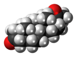
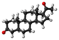
 Progesterone, a steroid hormone involved in the female menstrual cycle, pregnancy, and embryogenesis. |
 Medrogestone, a synthetic drug with similar effects as progesterone. |
 β-Sitosterol, a plant or phytosterol, with a fully branched hydrocarbon side chain at C-17, and an hydroxyl group at C-3. |
In addition to the ring scissions (cleavages), and expansions and contractions (cleavage and reclosing to a larger or smaller rings) noted in the IUPAC definition—all variations in the carbon-carbon bond framework—steroids can also vary:
- in the bond orders within the rings,
- in the number of methyl groups attached to the ring (and, when present, on the prominent side chain at C17),
- in the functional groups attached to the rings and side chain, and
- in the configuration of groups attached to the rings and chain.[1]:2–9
For instance, sterols such as cholesterol and lanosterol have an hydroxyl group attached at position C-3, while testosterone and progesterone have a carbonyl (oxo substituent) at C-3; of these examples, lanosterol alone has two methyl groups at C-4, and cholesterol with a C-5 to C-6 double bond differs from testosterone and progesterone, which have a C-4 to C-5 double bond.
Species distribution and function
The following are some of the common categories of steroids. In eukaryotes, steroids are found in the fungi, animals, and plants. Fungal steroids include the ergosterols.
The animal steroids include compounds of vertebrate and insect origin, in the latter case including ecdysteroids such as ecdysterone, which is involved in the control of molting in some species. Vertebrate examples include the steroid hormones and cholesterol, the latter of which is a structural component of cell membranes that is involved in determining the fluidity of cell membranes and is a principal constituent of plaques implicated in atherosclerosis. The steroid hormones include:
- the sex hormones that influence sex differences and support reproduction; these include androgens, estrogens, and progestagens;
- the corticosteroids, including the preponderance of synthetic steroid drugs, with natural product classes being the glucocorticoids that regulate many aspects of metabolism and immune function, and the mineralocorticoids that help maintain blood volume and control renal excretion of electrolytes; and
- the anabolic steroids, natural and synthetic, that interact with androgen receptors to increase muscle and bone synthesis, where in popular expressions, use of the term "steroids" may refer to anabolic steroids.
Plant steroids include steroidal alkaloids found in Solanaceae, the phytosterols, and the brassinosteroids (which include several plant hormones).
In prokaryotes, biosynthetic pathways exist both for producing the tetracyclic steroid framework (e.g., in mycobacteria)[6] — where its origin from eukaryotes is conjectured[7] — as well as the more common pentacyclic triterpinoid hopanoid framework.[8]
Types
Intact ring system
It is also possible to classify steroids based upon their chemical composition. One example of how MeSH performs this classification is available at the Wikipedia MeSH catalog. Examples from this classification include:


| Class | Examples | Number of carbon atoms |
|---|---|---|
| Cholestanes | cholesterol | 27 |
| Cholanes | cholic acid | 24 |
| Pregnanes | progesterone | 21 |
| Androstanes | testosterone | 19 |
| Estranes | estradiol | 18 |
The gonane (or steroid nucleus) is the parent (17-carbon tetracyclic) hydrocarbon molecule without any alkyl sidechains.[9]
Cleaved, contracted, and expanded rings
Secosteroids (L. seco, "to cut") are a subclass of steroidal compounds resulting, biosynthetically or conceptually, via scission (cleavage) of parent steroid rings, generally one of the four. Major secosteroid subclasses are defined by the steroid carbon atoms where this scission has taken place. For instance, the prototypical secosteroid cholecalciferol, vitamin D3 (shown), is in the important 9,10-secosteroid subclass, derived via cleavage between carbon atoms C-9 and C-10 of the steroid B-ring (similarly 5,6-secosteroids, 13,14-steroids, etc.).[10]
Norsteroids (nor-, L. norma, from "normal" in chemistry, indicating carbon removal)[11][12] and homosteroids (homo-, Gk. homos for same, indicating carbon addition)[13][12] are two structural subclasses of steroids formed via biosynthetic or bench chemistry steps, in the former case involving enzymic ring expansion/contraction reactions, and in the latter accomplished similarly (biomimetically) or, more often, through ring closures of acyclic precursors with more or fewer ring atoms than in the parent steroid framework.[14] The effect of these chemical operations on the rings is such that recognition of the parent tetracyclic steroid ring system can sometimes be challenging to the non-specialist. These two classes represent further unique classes of steroids with important biological activities and societal impacts.[14]
Combinations of these ring alterations are also possible and are known in nature. For instance, ewes that graze on corn lily ingest cyclopamine (shown) and veratramine, two of a sub-family of steroids where the C- and D-rings are contracted and expanded, respectively, via a biosynthetic migration of the original C-13 atom. Ingestion of these C-nor-D-homosteroids result in birth defects in progeny lambs: cyclopia in the case of cyclopamine and leg deformity with veratramine.[15] A further C-nor-D-homosteroid, nakiterpiosin, is excreted by Okinawan cyanobacteriosponges, Terpios hoshinota, leading to coral mortality from black coral disease.[16] Nakiterpiosin-type steroids are active against the signaling pathway involving the smoothened and hedgehog proteins, a pathway that is hyperactive in various cancers; Merck chemists established that C-13 atom migration could be achieved by "bench chemistry", and this biomimetic synthesis allows medicinal chemistry to proceed on this steroidal anticancer hypothesis.[15]
Biological significance
Steroid and their metabolites are frequently used signalling molecules. The most notable examples are the steroid hormones.
Steroids along with phospholipids function as components of cell membranes. Steroids such as cholesterol decrease membrane fluidity.[17]
Similar to lipids, steroids represent highly concentrated energy stores. However, steroids are not typically used as sources of energy. In mammals, they are normally metabolized and excreted.
Pharmacological actions
A number of drugs target the mevalonate pathway:
- Statins (used to reduce elevated cholesterol levels in patients)
- Bisphosphonates (used in treatment of various bone-degenerative diseases)
Biosynthesis and metabolism
The hundreds of distinct steroids found in animals, fungi, and plants are made either from lanosterol (in animals and fungi, see examples above) or from cycloartenol (in plants). Both lanosterol and cycloartenol are derived via cyclization of the triterpenoid squalene.[18]

Steroid biosynthesis is an anabolic metabolic pathway that produces steroids from simple precursors. A unique biosynthetic pathway is followed in animals compared to many other organisms, making the pathway a common target for antibiotics and other anti-infective drugs. In addition, steroid metabolism in humans is the target of cholesterol-lowering drugs such as statins.
In humans and other animals, the biosynthesis of steroids follows the mevalonate pathway that uses acetyl-CoA as building-blocks to form dimethylallyl pyrophosphate (DMAPP) and isopentenyl pyrophosphate (IPP).[19] In subsequent steps, DMAPP and IPP are joined to form geranyl pyrophosphate (GPP), which in turn is used to synthesize the steroid lanosterol. Further modifications of lanosterol into other steroids are classified steroidogenesis transformations.
Mevalonate pathway

The mevalonate pathway or HMG-CoA reductase pathway starts with acetyl-CoA and ends with dimethylallyl pyrophosphate (DMAPP) and isopentenyl pyrophosphate (IPP).
DMAPP and IPP in turn donate isoprene units, which are assembled and modified to form terpenes and isoprenoids,[20] which are a large class of lipids that include the carotenoids, and form the largest class of plant natural products.[21]
Here, the isoprene units are joined together to make squalene and then folded up and formed into a set of rings to make lanosterol.[22] Lanosterol can then be converted into other steroids such as cholesterol and ergosterol.[22][23]
Steroidogenesis

Steroidogenesis is the biological process by which steroids are generated from cholesterol and transformed into other steroids.[24] The pathways of steroidogenesis differ between different species.
Following is a list of the major classes of steroid hormones and some prominent members, with examples of major related functions:
- Progestogens:
- Progesterone, which regulates the cyclical changes of the endometrium of the uterus, and the maintenance of pregnancy
- Corticosteroids (Corticoids):
- Aldosterone (mineralocorticoids), which contributes to the regulation of blood pressure
- Cortisol (glucocorticoids), whose functions include acting as an immunosuppressant
- Androgens:
- Testosterone, which contributes to the development and maintenance of male secondary sex characteristics
- Estrogens:
- Estrogen, which contributes to the development and maintenance of female secondary sex characteristics
Locations of human steroidogenesis:
- Progestogens serve as precursors to all other human steroids – thus all human tissues which produce steroids must first convert cholesterol to pregnenolone. This conversion is the rate-limiting step of steroid synthesis, which occurs inside the mitochondrion of the respective tissue.[25]
- Corticosteroids are produced in the adrenal cortex.
- Estrogen and progesterone are made primarily in the ovary and in the placenta during pregnancy, and testosterone in the testes.
- Testosterone is also converted into estrogen to regulate the supply of each, in the bodies of both females and males.
- In addition, certain neurons and glia in the central nervous system (CNS) express the enzymes that are required for the local synthesis of pregnane neurosteroids, either de novo or from peripherally derived sources.
Regulation
Several key enzymes can be activated through DNA transcriptional regulation on activation of SREBP (sterol regulatory element-binding protein-1 and -2). This intracellular sensor detects low cholesterol levels and stimulates endogenous production by the HMG-CoA reductase pathway, as well as increasing lipoprotein uptake by up-regulating the LDL receptor. Regulation of this pathway is also achieved by controlling the rate of translation of the mRNA, degradation of reductase and phosphorylation.
Alternative pathways
In plants and bacteria, the non-mevalonate pathway uses pyruvate and glyceraldehyde 3-phosphate as substrates.[20][26]
Catabolism and excretion
Steroids are oxidized mainly by cytochrome P450 oxidase enzymes, such as CYP3A4. These reactions introduce oxygen into the steroid ring and allow the cholesterol structure to be broken up by other enzymes, to form bile acids as final products.[27] These bile acids can then be eliminated through secretion from the liver in the bile.[28] The expression of this oxidase gene can be upregulated by the steroid sensor PXR when there is a high blood concentration of steroids.[29]
Steroid hormones, lacking the side chain of cholesterol and bile acids, are typically hydroxylated at various ring positions and/or oxidized at the 17 position, then conjugted with sulfate or glucuronic acid and excreted in the urine.[30]
Isolation, structure determination, and methods of analysis
The isolation of steroids refers, depending on context, either to the isolation of the considerable quantities of pure chemical matter required for chemical structure elucidation, derivitzation/degradation chemistry, biological testing, and other research needs (generally milligrams to grams, but historically, often more),[31] or to the isolation of "analytical quantities" of the substance of interest, where the focus is on identification and quantitation of the substance (e.g., in biological tissue or fluid), and where the amount isolated depends on the analytical method applied (but is generally always sub-microgram in scale).[32] The methods of isolation applied toward achieving these two distinct scales of product are likewise distinct, but generally involve extraction, precipitation, adsorptions, chromatography, and sometimes crystallizations. In both cases, the isolated substance is purified to chemical homogeneity, i.e., specific combined separation and analytical methods such as LC-MS methods are chosen to be "orthogonal"—achieving their separations based on distinct modes of interaction between substance and isolating matrix—with the goal being detection of only a single species present in the purportedly pure sample. The expression structure determination refers to methods that are applied to determine the chemical structure of an isolated, pure steroid, a process that involves an array of chemical and physical methods that have changed markedly over the history of steroid research, but that have included NMR and small molecule crystallography.[1]:10–19 Methods of analysis include samplings of both of these prior areas, but especially analytical methods aimed at determining if a steroid is present in an analytical mixture, and determining its quantity in that medium.[32]
Chemical synthesis of steroids
Microbial transformations
Phytosterols, for instance, mixtures of soybean sterols, can be used as starting materials and converted into two kinds of steroid hormone intermediates through microbial transformation. Microbial catabolism of phytosterol sidechains yields either C-19 steroids, a precursor to most steroid hormones including sex hormones, or C-22 steroids, a precursor to adrenocortical hormones.[33][34]
Partial and total chemical synthesis
The chemical conversion of sapogenins to steroids—e.g., via the Marker degradation—is a method of partial synthesis that is a long-established alternative to microbial transformation of phytosterols to steroids, and underpinned Syntex efforts using the Mexican barbasco trade (harvesting and marketing large tubers of wild-growing plants, e.g., yams) to produce early synthetic steroids.
History
A number of Nobel Prizes have been awarded for research involving steroids. These prizes include:
- 1927 (Chemistry) Heinrich Otto Wieland – constitution of the bile acids, sterols, and their connection with the vitamins[35]
- 1928 (Chemistry) Adolf Otto Reinhold Windaus – constitution of the sterols and their connection with the vitamins[36]
- 1939 (Chemistry) Adolf Butenandt and Leopold Ruzicka – isolation and structural studies of steroid sex hormones, and related studies on higher terpenes[37]
- 1950 (Physiology or Medicine) Edward Calvin Kendall, Tadeus Reichstein, Philip Hench – on the structure and biological effects of adrenal hormones[38]
- 1965 (Chemistry) Robert Burns Woodward, in part for the synthesis of cholesterol, cortisone, and lanosterol[39]
- 1969 (Chemistry) Derek Barton, Odd Hassel, development of the concept of conformation and its application in chemistry, where a specific important emphasis was on the conformation of the "Steroid Nucleus"[40]
- 1975 (Chemistry) Vladimir Prelog, in part for developing methods to determine the stereochemical course of cholesterol biosynthesis from mevalonic acid via squalene[41]
See also
References
- ↑ 1.0 1.1 1.2 Lednicer D (2011). Steroid Chemistry at a Glance. Hoboken: Wiley. ISBN 978-0-470-66084-3.
- ↑ Rhen T, Cidlowski JA (2005). "Antiinflammatory action of glucocorticoids — new mechanisms for old drugs" (PDF). N. Engl. J. Med. 353 (16): 1711–23. doi:10.1056/NEJMra050541. PMID 16236742.
- ↑ 3.0 3.1 Moss GP (1989). "Nomenclature of Steroids (Recommendations 1989)". Pure & Appl. Chem. 61 (10): 1783–1822. doi:10.1351/pac198961101783. PDF "IUPAC-IUB Joint Commission on Biochemical Nomenclature (JCBN). The nomenclature of steroids. Recommendations 1989". Eur. J. Biochem. 186 (3): 429–58. December 1989. doi:10.1111/j.1432-1033.1989.tb15228.x. PMID 2606099.
- ↑ 4.0 4.1 4.2 G.P. Moss and the Working Party of the IUPAC-IUB Joint Commission on Biochemical Nomenclature, "The Nomenclature of Steroids", hosted at Queen Mary University of London, Section 3S-1 (esp. 3S-1.4, incl. note 4) See and , accessed 10 May 2014. Also available from same authors at Pure Appl. Chem. 1989, 61, 1783-1822 (esp. p. 1785f), or R.A. Hill, D.N. Kirk, H.L.J. Makin, H.L.J. & G.M. Murphy, 1991, "Dictionary of Steroids" London:Chapman and Hall, pp. xxx-lix. The Working Party of the IUPAC-IUB JCBN were P. Karlson (chairman), J.R. Bull, K. Engel, J. Fried†, H.W. Kircher, K.L. Loening, G.P. Moss, G. Popják and M.R. Uskokovic.
- ↑ PubChem 130801; 219-08-9 cyclopentaphenanthrene
- ↑ Bode HB, Zeggel B, Silakowski B, Wenzel SC, Reichenbach H, Müller R (2003). "Steroid biosynthesis in prokaryotes: identification of myxobacterial steroids and cloning of the first bacterial 2,3(S)-oxidosqualene cyclase from the myxobacterium Stigmatella aurantiaca". Mol. Microbiol. 47 (2): 471–81. doi:10.1046/j.1365-2958.2003.03309.x. PMID 12519197.
- ↑ Desmond E, Gribaldo S (2009). "Phylogenomics of sterol synthesis: insights into the origin, evolution, and diversity of a key eukaryotic feature". Genome Biol Evol 1: 364–81. doi:10.1093/gbe/evp036. PMC 2817430. PMID 20333205.
- ↑ Siedenburg G, Jendrossek D (2011). "Squalene-hopene cyclases". Appl. Environ. Microbiol. 77 (12): 3905–15. doi:10.1128/AEM.00300-11. PMC 3131620. PMID 21531832.
- ↑ Edgren RA, Stanczyk FZ (December 1999). "Nomenclature of the gonane progestins". Contraception 60 (6): 313. doi:10.1016/S0010-7824(99)00101-8. PMID 10715364.
- ↑ Hanson JR (June 2010). "Steroids: partial synthesis in medicinal chemistry". Nat Prod Rep 27 (6): 887–99. doi:10.1039/c001262a. PMID 20424788.
- ↑ International Union of Pure and Applied Chemistry (IUPAC), 1999, "RF-4.1 Removal of Skeletal Atoms," in "RF-4. Skeletal Modifications" in Revised Section F: Natural Products and Related Compounds (IUPAC Recommendations 1999), see , accessed 20 May 2014.
- ↑ 12.0 12.1 See also IUPAC, 1976, "Nomenclature of Organic Chemistry: Section F - Natural Products and Related Compounds, Recommendations 1976", IUPAC Information Bulletin Appendices on Tentative Nomenclature, Symbols, Units, and Standards, No. 53, December, 1976, also in Eur. J. Biochem. 1978, 86, 1-8.
- ↑ International Union of Pure and Applied Chemistry (IUPAC), 1999, "RF-4.2. Addition of Skeletal Atoms," in "RF-4. Skeletal Modifications" in Revised Section F: Natural Products and Related Compounds (IUPAC Recommendations 1999), see , accessed 20 May 2014.
- ↑ 14.0 14.1 János Wölfling, 2007, "Recent developments in the isolation and synthesis of D-homosteroids and related compounds" (Issue in Honor of Prof. Lutz F. Tietze), ARKIVOC (v) 210-230. See , accessed 20 May 2014.
- ↑ 15.0 15.1 Here and following, see Shuanhu Gao & Chio Chen, 2012, "Nakiterpiosin", in Total Synthesis of Natural Products: At the Frontiers of Organic Chemistry (Jie Jack Li & E.J. Corey, eds.), Berlin:Springer, pp. 25-38, esp. 25-28, e.g., , accessed 20 May 2014.
- ↑ D. Uemura, M. Kita, H. Arimoto & M. Kitamura, 2009, "Recent aspects of chemical ecology: Natural toxins, coral communities, and symbiotic relationships," Pure Appl. Chem., 81(6), 1093–1111, esp. 1101. DOI 10.1351/PAC-CON-08-08-12. See , accessed 20 May 2014.
- ↑ Sadava D, Hillis DM, Heller HC, Berenbaum MR (2011). Life: The Science of Biology 9th Edition. San Francisco: Freeman. pp. 105–114. ISBN 1-4292-4646-4.
- ↑ "Lanosterol biosynthesis". Recommendations on Biochemical & Organic Nomenclature, Symbols & Terminology. International Union Of Biochemistry And Molecular Biology.
- ↑ Grochowski L, Xu H, White R (2006). "Methanocaldococcus jannaschii uses a modified mevalonate pathway for biosynthesis of isopentenyl diphosphate". J Bacteriol 188 (9): 3192–8. doi:10.1128/JB.188.9.3192-3198.2006. PMC 1447442. PMID 16621811.
- ↑ 20.0 20.1 Kuzuyama T, Seto H (2003). "Diversity of the biosynthesis of the isoprene units". Nat Prod Rep 20 (2): 171–83. doi:10.1039/b109860h. PMID 12735695.
- ↑ Dubey V, Bhalla R, Luthra R (2003). "An overview of the non-mevalonate pathway for terpenoid biosynthesis in plants" (PDF). J Biosci 28 (5): 637–46. doi:10.1007/BF02703339. PMID 14517367.
- ↑ 22.0 22.1 Schroepfer G (1981). "Sterol biosynthesis". Annu Rev Biochem 50: 585–621. doi:10.1146/annurev.bi.50.070181.003101. PMID 7023367.
- ↑ Lees N, Skaggs B, Kirsch D, Bard M (1995). "Cloning of the late genes in the ergosterol biosynthetic pathway of Saccharomyces cerevisiae—a review". Lipids 30 (3): 221–6. doi:10.1007/BF02537824. PMID 7791529.
- ↑ Hanukoglu I (Dec 1992). "Steroidogenic enzymes: structure, function, and role in regulation of steroid hormone biosynthesis.". J Steroid Biochem Mol Biol 43 (8): 779–804. doi:10.1016/0960-0760(92)90307-5. PMID 22217824.
- ↑ Rossier MF (2006). "T channels and steroid biosynthesis: in search of a link with mitochondria". Cell Calcium. 40 (2): 155–64. doi:10.1016/j.ceca.2006.04.020. PMID 16759697.
- ↑ Lichtenthaler H (1999). "The 1-Dideoxy-D-xylulose-5-phosphate pathway of isoprenoid biosynthesis in plants". Annu Rev Plant Physiol Plant Mol Biol 50: 47–65. doi:10.1146/annurev.arplant.50.1.47. PMID 15012203.
- ↑ Pikuleva IA (2006). "Cytochrome P450s and cholesterol homeostasis". Pharmacol. Ther. 112 (3): 761–73. doi:10.1016/j.pharmthera.2006.05.014. PMID 16872679.
- ↑ Zollner G, Marschall HU, Wagner M, Trauner M (2006). "Role of nuclear receptors in the adaptive response to bile acids and cholestasis: pathogenetic and therapeutic considerations". Mol. Pharm. 3 (3): 231–51. doi:10.1021/mp060010s. PMID 16749856.
- ↑ Kliewer S, Goodwin B, Willson T (2002). "The nuclear pregnane X receptor: a key regulator of xenobiotic metabolism". Endocr. Rev. 23 (5): 687–702. doi:10.1210/er.2001-0038. PMID 12372848.
- ↑ Thomas Steimer "Steroid Hormone Metabolism",Geneva Foundation for Medical Education and Research, 2, ch. du Petit Bel-Air, 1225 Chêne-Bourg, Geneva, Switzerland
- ↑ American Chemical Society, International Historic Chemical Landmark, 1999, "Russell Marker and the Mexican Steroid Hormone Industry," see , accessed 10 May 2104.
- ↑ 32.0 32.1 Makin HLJ, Gower DB (2010). Steroid analysis. Dordrecht; New York: Springer. ISBN 978-1-4020-9774-4.
- ↑ Conner AH, Nagaoka M, Rowe JW, Perlman D (August 1976). "Microbial conversion of tall oil sterols to C19 steroids" (PDF). Appl. Environ. Microbiol. 32 (2): 310–1. PMC 170056. PMID 987752.
- ↑ Wang F-Q, Yao K, Wei D-Z. "From Soybean Phytosterols to Steroid Hormones, Soybean and Health". In El-Shemy H. Soybean and Health. InTech. doi:10.5772/18808. ISBN 978-953-307-535-8.
- ↑ "The Nobel Prize in Chemistry 1927". The Nobel Foundation.
- ↑ "The Nobel Prize in Chemistry 1928". The Nobel Foundation.
- ↑ "The Nobel Prize in Chemistry 1939". The Nobel Foundation.
- ↑ "The Nobel Prize in Physiology or Medicine 1950". The Nobel Foundation.
- ↑ "The Nobel Prize in Chemistry 1965". The Nobel Foundation.
- ↑ "The Nobel Prize in Chemistry 1969". The Nobel Foundation.
- ↑ "The Nobel Prize in Chemistry 1975". The Nobel Foundation.
Further reading
- Lednicer D (2011). Steroid Chemistry at a Glance. Hoboken: Wiley. doi:10.1002/9780470973639. ISBN 978-0-470-66085-0.
- Yoder RA, Johnston JN (December 2005). "A case study in biomimetic total synthesis: polyolefin carbocyclizations to terpenes and steroids". Chem. Rev. 105 (12): 4730–56. doi:10.1021/cr040623. PMC 2575671. PMID 16351060. – review of the history of steroid synthesis, especially biomimetic
- Han, Thang S.; Walker, Brian R.; Arlt, Wiebke; Ross, Richard J. (17 December 2013). "Treatment and health outcomes in adults with congenital adrenal hyperplasia". Nature Reviews Endocrinology 10 (2): 115–124. doi:10.1038/nrendo.2013.239. PMID 24342885Figure 2: The adrenal steroidogenesis pathway.
External links
- Moss GP; IUPAC-IUB Joint Commission on Biochemical Nomenclature (JCBN). "The Nomenclature of Steroids Home Page". Queen Mary University of London.
- Bowen RA (October 20, 2001). "Steroidogenesis". Pathophysiology of the Endocrine System. Colorado State University.
| ||||||||||||||||||||||||||||||||||||||||||||||||||||||||||||||||||||||||||||||||||||||||||||||||||||||||||||||||||||||||||||
| ||||||||||||||||||||||||||||||||||||||||||||||||||||||||||||||||||||||||||||||||||||||||||||||||||||||||||||||||||||||
| ||||||||||||||||||||||||||||||||||||||||||||||||||||||||||||||||||||||||||||||||||||||||||||||