Right atrium
| Right atrium | |
|---|---|
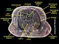
.svg.png) Anterior (frontal) view of the opened heart. White arrows indicate normal blood flow. | |
 Interior of right side of heart. | |
| Details | |
| Latin | atrium dextrum |
| Identifiers | |
| Gray's | p.528 |
| MeSH | A07.541.358 |
| Dorlands /Elsevier | a_71/12167861 |
| TA | A12.1.01.001 |
| FMA | 7096 |
| Anatomical terminology | |
The right atrium (in older texts termed the "right auricle", which now means the right atrial appendage) is one of four chambers (two atria and two ventricles) in the hearts of mammals (including humans) and archosaurs (which include birds and crocodilians). It receives deoxygenated blood from the superior and inferior venae cavae, the coronary sinus, and the anterior and smallest cardiac veins, and pumps it into the right ventricle through the tricuspid valve. Attached to the right atrium is the right atrial appendage.
Structure
- Sinus venarum smooth-walled portion that surrounds the opening of the superior and inferior vena cava and the coronary sinus.
a.This region is the adult remnant of the embryonic sinus venosus. - Openings of three vessels:
a. Superior Vena Cava
b. Inferior Vena Cava
c. Coronary Sinus - Pectinate muscles (musculi pectinati): muscular wall of the atria
- Right atrial appendage pouch-like extension of the muscular part (pectinate muscles) of the right atrium
- Crista terminalis a ridge separating the muscular and smooth walled parts of the right atrium
- Interatrial septum: a barrier separating the right atrium from the left atrium
- Fossa ovalis: remnant of the closure of an opening that was present within the interatrial septum of the fetal heart (normal) where it is called the foramen ovale. It is a depression that looks like "thumb print" in the interatrial wall.
- Tricuspid valve: its superior surface is from the right atrium.
Nodes
The sinoatrial node (SAN) is located within this chamber next to the vena cava. This is a group of pacemaker cells which spontaneously depolarize to create an action potential. The cardiac action potential then spreads across both atria causing them to contract, forcing the blood they hold into their corresponding ventricles.
Development
During embryogenesis the septum in the right atrium has an opening, the foramen ovale which provides access to the left atrium; this connects the two chambers, which is essential for fetal blood circulation. At birth the foramen ovale closes and leaves a depression (the fossa ovalis) in the atrial wall. When the first breath is taken, fetal blood flow is reversed and now travels through the lungs, no longer requiring the foramen ovale.
In some cases, the foramen ovale fails to close and is present in 20% of the general population; This is known as a patent foramen ovale. However, it is mostly unproblematic.
Within the fetal right atrium, blood from the inferior vena cava and the superior vena cava flow in separate streams to different locations in the heart, and this has been reported to occur through the Coandă effect.[1]
Function
Atria facilitate circulation primarily by allowing uninterrupted venous flow to the heart, preventing the inertia of interrupted venous flow that would otherwise occur at each ventricular systole.[2] The right atrium is composed of a smooth intercaval part and a muscular part (formed by pectinate muscles) separated by an internal ridge called the crista terminalis and an external groove (sulcus terminalis). The muscular part is derived from the primitive atrium while the smooth part is formed from the embryological sinus venosus.
Additional images
-

Heart right anatomy
-
Right atrium
References
- ↑ Ashrafian H. The Coanda effect and preferential right atrial streaming. Chest. 2006 Jul;130(1):300.
- ↑ Anderson, RM. The Gross Physiology of the Cardiovascular System (2nd ed., 2012). See "Chapter 1: Normal Physiology."