Ribonuclease H
| ribonuclease H | |||||||||
|---|---|---|---|---|---|---|---|---|---|
 | |||||||||
| Identifiers | |||||||||
| EC number | 3.1.26.4 | ||||||||
| CAS number | 9050-76-4 | ||||||||
| Databases | |||||||||
| IntEnz | IntEnz view | ||||||||
| BRENDA | BRENDA entry | ||||||||
| ExPASy | NiceZyme view | ||||||||
| KEGG | KEGG entry | ||||||||
| MetaCyc | metabolic pathway | ||||||||
| PRIAM | profile | ||||||||
| PDB structures | RCSB PDB PDBe PDBsum | ||||||||
| Gene Ontology | AmiGO / EGO | ||||||||
| |||||||||
| retroviral ribonuclease H | |||||||||
|---|---|---|---|---|---|---|---|---|---|
| Identifiers | |||||||||
| EC number | 3.1.26.13 | ||||||||
| Databases | |||||||||
| IntEnz | IntEnz view | ||||||||
| BRENDA | BRENDA entry | ||||||||
| ExPASy | NiceZyme view | ||||||||
| KEGG | KEGG entry | ||||||||
| MetaCyc | metabolic pathway | ||||||||
| PRIAM | profile | ||||||||
| PDB structures | RCSB PDB PDBe PDBsum | ||||||||
| |||||||||
Ribonuclease H (RNase H) is a family of non-sequence-specific endonucleases that catalyze the cleavage of RNA via a hydrolytic mechanism. Members of the RNase H family can be found in nearly all organisms, from bacteria to archaea to eukaryotes.
RNase H’s ribonuclease activity cleaves the 3’-O-P bond of RNA in a DNA/RNA duplex substrate to produce 3’-hydroxyl and 5‘-phosphate terminated products. In DNA replication, RNase H is responsible for removing the RNA primer, allowing completion of the newly synthesized DNA.
Structure
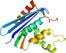
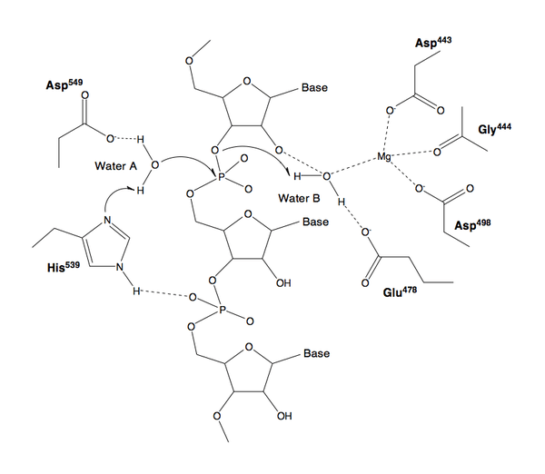
The 3-D structure of RNase H commonly consists of a 5-stranded β-sheet surrounded by a distribution of α-helices.[2] In some RNase H, such as the one found in HIV-1, the enzyme is missing one of the helices known as the C-helix, a positively charged α-helix whose protrusive shape increases substrate binding capacity.[3] The active site of the enzyme is centered around a conserved DEDD motif (composed of residues: D443, E478, D498, and D549) which performs the hydrolysis of the RNA substrate.[4] A magnesium ion is commonly used as a cofactor during the hydrolysis step.[5] It is also a potential but unconfirmed mechanism in which multiple ions are necessary to perform the hydrolysis.[6] The enzyme also contains a nucleic acid binding cleft about 60 Å in length that can encompass a region of 18 bound RNA/DNA base pairs.
Function
Because RNase H specifically degrades only the RNA in RNA:DNA hybrids, it is commonly used in molecular biology to destroy the RNA template after first-strand complementary DNA (cDNA) synthesis by reverse transcription, as well as in procedures such as nuclease protection assays. RNase H can also be used to degrade specific RNA strands when the cDNA oligonucleotide is hybridized, such as the removal of the polyadenine tail from mRNA hybridized to oligo(dT), or the destruction of a chosen non-coding RNA inside or outside the living cell. To terminate the reaction, a chelator, such as EDTA, is often added to sequester the required metal ions in the reaction mixture.
Mechanism
Genes
The following human genes encode proteins with RNase H activity:
Role in Disease
Mutations in the RNASE2 genes are associated with the rare human genetic disorder known as Aicardi–Goutieres syndrome,[7] which manifests as neurological and dermatological symptoms at an early age.[8]
Retroviral RNase H, a part of the viral reverse transcriptase enzyme, is an important pharmaceutical target, as it is absolutely necessary for the proliferation of retroviruses, such as HIV and murine leukemia virus.[9][10] Inhibitors of this enzyme could therefore provide new drugs against diseases like AIDS. While not an effective treatment option, incorporation of 6-deoxythioguanosine has been shown to inhibit RNase H cleavage of the DNA/RNA complex.[11]
HIV-1
Within human immunodeficiency virus type 1 (HIV-1), RNase H exists as a domain in the heterodimeric HIV-1 reverse transcriptase enzyme.[12] HIV-1 carries out reverse transcription, a process that produces new double-stranded DNA from the viral genome's single-stranded RNA. During DNA synthesis, a DNA/RNA hybrid is formed as a replication intermediate and must be cleaved by RNase H before the process can continue. RNase H performs three types of cleaving actions: non-specific degradation of the (+)-strand RNA genome, specific removal of the (-)-strand tRNA primer, and removal of the (+)-strand purine-rich polypurine tract (PPT) primer.[13] RNase H plays a role in the priming of the (+)-strand, but not in the conventional method of synthesizing a new primer sequence. Rather RNase H creates a "primer" from the PPT that is resistant to RNase H cleavage. By removing all bases but the PPT, the PPT is used as a marker for the end of the U3 region of its long terminal repeat.[14]
References
- ↑ PDB 1JL1; Goedken ER, Marqusee S (December 2001). "Native-state energetics of a thermostabilized variant of ribonuclease HI". J. Mol. Biol. 314 (4): 863–871. doi:10.1006/jmbi.2001.5184. PMID 11734003.
- ↑ Schmitt TJ, Clark JE, Knotts TA (December 2009). "Thermal and mechanical multistate folding of ribonuclease H". J Chem Phys 131 (23): 235101. doi:10.1063/1.3270167. PMID 20025349.
- ↑ Schultz SJ, Champoux JJ (June 2008). "RNase H activity: structure, specificity, and function in reverse transcription". Virus Res. 134 (1–2): 86–103. doi:10.1016/j.virusres.2007.12.007. PMC 2464458. PMID 18261820.
- ↑ Tadokoro T, Kanaya S (March 2009). "Ribonuclease H: molecular diversities, substrate binding domains, and catalytic mechanism of the prokaryotic enzymes". FEBS J. 276 (6): 1482–1493. doi:10.1111/j.1742-4658.2009.06907.x. PMID 19228197.
- ↑ Davies JF, Hostomska Z, Hostomsky Z, Jordan SR, Matthews DA (April 1991). "Crystal structure of the ribonuclease H domain of HIV-1 reverse transcriptase". Science 252 (5002): 88–95. doi:10.1126/science.1707186. PMID 1707186.
- ↑ Klumpp K; Hang JQ; Rajendran S et al. (December 2003). "Two-metal ion mechanism of RNA cleavage by HIV RNase H and mechanism-based design of selective HIV RNase H inhibitors". Nucleic Acids Res. 31 (23): 6852–9. doi:10.1093/nar/gkg881. PMC 290251. PMID 14627818.
- ↑ Crow, Y. J.; Leitch, A.; Hayward, B. E.; Garner, A.; Parmar, R.; Griffith, E.; Ali, M.; Semple, C.; Aicardi, J.; Babul-Hirji, R.; Baumann, C.; Baxter, P.; Bertini, E.; Chandler, K. E.; Chitayat, D.; Cau, D.; Déry, C.; Fazzi, E.; Goizet, C.; King, M. D.; Klepper, J.; Lacombe, D.; Lanzi, G.; Lyall, H.; Martínez-Frías, M. A. L.; Mathieu, M. L.; McKeown, C.; Monier, A.; Oade, Y.; Quarrell, O. W. (2006). "Mutations in genes encoding ribonuclease H2 subunits cause Aicardi-Goutières syndrome and mimic congenital viral brain infection". Nature Genetics 38 (8): 910–916. doi:10.1038/ng1842. PMID 16845400.
- ↑ Orcesi, S; La Piana, R; Fazzi, E (2009). "Aicardi-Goutieres syndrome". British Medical Bulletin 89: 183–201. doi:10.1093/bmb/ldn049. PMID 19129251.
- ↑ Mizuno M, Yasukawa K, Inouye K (February 2010). "Insight into the mechanism of the stabilization of moloney murine leukaemia virus reverse transcriptase by eliminating RNase H activity". Biosci. Biotechnol. Biochem. 74 (2): 440–2. doi:10.1271/bbb.90777. PMID 20139597.
- ↑ Coté ML, Roth MJ (June 2008). "Murine leukemia virus reverse transcriptase: structural comparison with HIV-1 reverse transcriptase". Virus Res. 134 (1–2): 186–202. doi:10.1016/j.virusres.2008.01.001. PMC 2443788. PMID 18294720.
- ↑ Krynetskaia NF, Krynetski EY, Evans WE (October 1999). "Human RNase H-mediated RNA cleavage from DNA-RNA duplexes is inhibited by 6-deoxythioguanosine incorporation into DNA". Mol. Pharmacol. 56 (4): 841–8. PMID 10496969.
- ↑ http://www.mdpi.com/1999-4915/2/4/900/pdf
- ↑ Klarmann GJ, Hawkins ME, Le Grice SF (2002). "Uncovering the complexities of retroviral ribonuclease H reveals its potential as a therapeutic target". AIDS Rev 4 (4): 183–94. PMID 12555693.
- ↑ Beilhartz GL, Götte M (March 2010). "HIV-1 Ribonuclease H: Structure, Catalytic Mechanism and Inhibitors". Viruses 1 (2): 900–926.
External links
- GeneReviews/NCBI/NIH/UW entry on Aicardi-Goutières Syndrome
- RNase H at the US National Library of Medicine Medical Subject Headings (MeSH)
| |||||||||||||||||||||||||||||||||||||||||||||||||||||||
| ||||||||||||||||||||||||||||||||||||||||||||||||