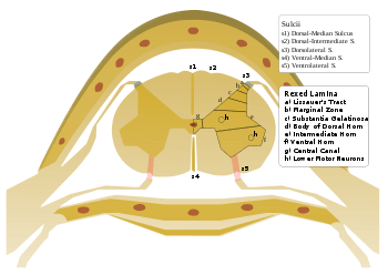
Rexed laminae

Medulla spinalis - Substantia grisea

Rexed lamina
The Rexed laminae comprise a system of ten layers of grey matter (I-X), identified in the early 1950s by Bror Rexed to label portions of the grey columns of the spinal cord. [1][2]
Similar to Brodmann areas, they are defined by their cellular structure rather than by their location, but the location still remains reasonably consistent.
Laminae
- Posterior/dorsal horn: I-VI
- Lamina I: marginal nucleus of spinal cord or posteromarginal nucleus
- Lamina II: substantia gelatinosa of Rolando
- Laminae III and IV: nucleus proprius
- Lamina V: Neck of the dorsal horn
- Lamina VI: Base of the dorsal horn,
- Intermediate zone: VII and X
- Lamina VII: intermediomedial nucleus, intermediolateral nucleus, nucleus dorsalis of Clarke in the thoracic and upper lumbar region[3]
- Lamina X: central gray matter i.e. neurons bordering Central canal; Grisea Centralis[3]
- Anterior/ventral horn: VIII-IX
- Lamina VIII: motor interneurons; Commissural nucleus[3]
- Lamina IX: lateral (in limb regions) and medial motor neurons, also phrenic and spinal accessory nuclei at cervical levels, and Onuf's nucleus in the sacral region
- Centrally
- Lamina X: Central Zone, grey matter surrounding the central canal
References
- ↑ Rexed B (1952). "The cytoarchitectonic organization of the spinal cord in the cat.". J Comp Neurol 96 (3): 414–95. doi:10.1002/cne.900960303. PMID 14946260.
- ↑ Rexed B (1954). "A cytoarchitectonic atlas of the spinal cord in the cat.". J Comp Neurol 100 (2): 297–379. doi:10.1002/cne.901000205. PMID 13163236.
- ↑ 3.0 3.1 3.2 Blumenfeld, Hal (2010). Neuroanatomy through Clinical Cases. Sunderland, MA: Sinauer Associates.
| ||||||||||||||||||||||||||||||||||||||||||||||||||||||||||||||||||||||||||||