Reticulocyte
| Reticulocyte | |
|---|---|
 Reticulocytes | |
 Erythrocytes (mature cells) | |
| Details | |
| Latin | reticulocytus |
| Identifiers | |
| Code | TH H2.00.04.1.01007 |
| Anatomical terminology | |
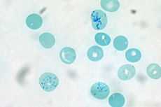
Reticulocytes are immature red blood cells, typically composing about 1% of the red cells in the human body. Reticulocytes develop and mature in the bone marrow and then circulate for about a day in the blood stream before developing into mature red blood cells. Like mature red blood cells, in mammals reticulocytes do not have a cell nucleus. They are called reticulocytes because of a reticular (mesh-like) network of ribosomal RNA that becomes visible under a microscope with certain stains such as new methylene blue.
Reticulocytes as a clinical indicator
To accurately measure reticulocyte counts, automated counters use a combination of laser excitation, detectors and a fluorescent dye that marks RNA and DNA (such as thiazole orange or polymethine).[1] Reticulocytes can be distinguished from other circulating cells because they emit a signal that is neither strong (like lymphocytes) nor weak (like red blood cells).[2]
Reticulocytes appear slightly bluer than other red cells when looked at with the normal Romanowsky stain. Reticulocytes are also relatively large, a characteristic that is described by the MCV (mean corpuscular volume).
The normal fraction of reticulocytes in the blood depends on the clinical situation but is usually 0.5% to 2.5% in adults and 2% to 6% in infants. A reticulocyte percentage that is higher than "normal" can be a sign of anemia, but this depends on the health of a person's bone marrow. Calculating the reticulocyte production index is an important step in understanding whether or not the reticulocyte count is appropriate to the situation. This is often a more important question than whether the percentage is in the normal range; for instance, if someone is anemic but has a reticulocyte percentage of only 1%, the bone marrow is likely not producing new blood cells at a rate that will correct the anemia.
The number of reticulocytes is a good indicator of bone marrow activity because it represents recent production and allows for the determination of reticulocyte count and the reticulocyte production index. These values can be used to determine whether a production problem is contributing to the anemia and can also be used to monitor the progress of treatment for anemia.
When there is an increased production of red blood cells to overcome chronic or severe loss of mature red blood cells, such as in a haemolytic anemia, people often have a markedly high number and percentage of reticulocytes. A very high number of reticulocytes in the blood can be described as reticulocytosis.
Abnormally low numbers of reticulocytes can be attributed to chemotherapy, aplastic anemia, pernicious anemia, bone marrow malignancies, problems of erythropoietin production, various vitamin or mineral deficiencies (B9, B12, iron), disease states (anemia of chronic disease) and other causes of anemia due to poor RBC production.
Research
Reticulocytes are a valuable tool for biologists who study protein translation. Reticulocytes are unusual among cells in that they contain all of the machinery necessary to translate proteins but lack a nucleus. Since a cell's nucleus contains many components that make studying translation difficult, these cells are quite useful. Scientists can collect reticulocytes from animals such as rabbits and extract the mRNA and translation enzymes to study protein translation in a cell-free, in vitro system, allowing greater control over the environment in which proteins are being synthesized.[3]
References
- ↑ Davis BH, Bigelow NC (1994). "Reticulocyte analysis and reticulocute maturity index". In Darzynkiewicz Z, Crissman HA (eds.). Flow cytometry. Methods in Cell Biology 42. San Diego: Academic Press. pp. 263–74. ISBN 0-12-203052-4.
- ↑ http://www.medicaldesign.com/articles/ID/532
- ↑ http://www.invitrogen.com/site/us/en/home/References/Ambion-Tech-Support/large-scale-transcription/general-articles/the-basics-in-vitro-translation.html
External links
- For more information on Reticulocyte Count
- ‹The template EMedicineDictionary is being considered for deletion.› Reticulocyte at eMedicine Dictionary
| ||||||||||||||||||||||||||||||||||||||||||||||||||||||||||||||||||||||
| ||||||||||||||||||||||||||||||||||||||||||||||||||||||||