Renal calyx
| Renal calyx | ||||
|---|---|---|---|---|
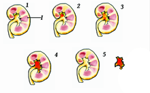
 Kidney, with major and minor calyces labeled near bottom. | ||||

| ||||
| Details | ||||
| Latin | calices renales | |||
| Precursor | Ureteric bud | |||
| Identifiers | ||||
| Gray's | p.1225 | |||
| MeSH | A05.810.453.537.503 | |||
| Dorlands /Elsevier | c_03/12206023 | |||
| Anatomical terminology | ||||
The renal calyces are chambers of the kidney through which urine passes. The minor calyces surround the apex of the renal pyramids. Urine formed in the kidney passes through a renal papilla at the apex into the minor calyx; two or three minor calyces converge to form a major calyx, through which urine passes before continuing through the renal pelvis into the ureter.
Function
Peristalsis of the smooth muscle originating in pace-maker cells originating in the walls of the calyces propels urine through the renal pelvis and ureters to the bladder.
Clinical significance
Example of a "staghorn" kidney stone projecting into the renal calyces
A "Staghorn calculi" is a kidney stone that may extend into the renal calyces.
A renal diverticulum is diverticulum of renal calyces.[1][2]
See also
References
- ↑ Krzeski, T; Witeska, A; Borówka, A; Pypno, W (September 1981). "Diverticula of renal calyces". International Urology and Nephrology 13 (3): 231–235. doi:10.1007/BF02082420.
- ↑ Chong, TW; Bui, MH; Fuchs, GJ (Nov 2000). "Calyceal diverticula. Ureteroscopic management.". The Urologic clinics of North America 27 (4): 647–54. doi:10.1016/s0094-0143(05)70114-2. PMID 11098763.
External links
- Anatomy photo:40:06-0108 at the SUNY Downstate Medical Center - "Posterior Abdominal Wall: Internal Structure of a Kidney"
- Anatomy photo:40:06-0109 at the SUNY Downstate Medical Center - "Posterior Abdominal Wall: Internal Structure of a Kidney"
- Histology image: 15901loa — Histology Learning System at Boston University - "Urinary System: neonatal kidney"
- posteriorabdomen at The Anatomy Lesson by Wesley Norman (Georgetown University) (renalpelvis)
- Diagram at bway.net
| ||||||||||||||||||||||||||||||||||||||||||||||||||||||||||||||