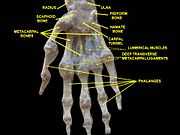
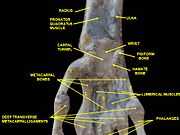
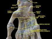
The radius or radial bone is one of the two large bones of the forearm, the other being the ulna. It extends from the lateral side of the elbow to the thumb side of the wrist and runs parallel to the ulna, which exceeds it in length and size. It is a long bone, prism-shaped and slightly curved longitudinally. The radius articulates with the capitulum of the humerus, and with the ulna at two points: the radial notch (lower) and the head (upper part) of the ulna. The corresponding bone in the lower leg is the tibia.
The word radius is Latin for "ray". In the context of the radius bone, a ray can be thought of rotating around an axis line extending diagonally from center of capitulum to the center of distal ulna. While the ulna is the major contributor to the elbow joint, the radius primarily contributes to the wrist joint.[1]
The radius is named so because the radius (bone) acts like the radius (of a circle). It rotates around the ulna and the far end (where it joins to the bones of the hand), known as the styloid process of the radius, is the distance from the ulna (center of the circle) to the edge of the radius (the circle). The ulna acts as the center point to the circle because when the arm is rotated the ulna does not move.
Structure
The long narrow medullary cavity is enclosed in a strong wall of compact bone. It is thickest along the interosseous border and thinnest at the extremities, save over the cup-shaped articular surface (fovea) of the head.
The trabeculae of the spongy tissue are somewhat arched at the upper end and pass upward from the compact layer of the shaft to the fovea capituli (the humerus's cup-shaped articulatory notch); they are crossed by others parallel to the surface of the fovea. The arrangement at the lower end is somewhat similar. It is missing in radial aplasia.
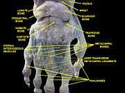
The radius has a body and two extremities. The upper extremity of the radius consists of a somewhat cylindrical head articulating with the ulna and the humerus, a neck, and a double tuberosity. The body of the radius is self-explanatory, and the lower extremity of the radius is roughly quadrilateral in shape, with articular surfaces for the ulna, scaphoid and lunate bones. The distal end of the radius forms two palpable points, radially the styloid process and Lister's tubercle on the ulnar side. Along with the proximal and distal radioulnar articulations, an interosseous membrane originates medially along the length of the body of the radius to attach the radius to the ulna.[2]
Muscle attachments
The biceps muscle inserts on the radial tuberosity of the upper extremity of the bone. The upper third of the body of the bone attaches to the supinator, the flexor digitorum superficialis, and the flexor pollicis longus muscles.
The middle third of the body attaches to the extensor ossis metacarpi pollicis, extensor primi internodii pollicis, and the pronator teres muscles.
The lower quarter of the body attaches to the pronator quadratus muscle and the tendon of the supinator longus.
In other animals
In four-legged animals, the radius is the main load-bearing bone of the lower forelimb. Its structure is similar in most terrestrial tetrapods, but it may be fused with the ulna in some mammals (such as horses) and reduced or modified in animals with flippers or vestigial forelimbs.[3]
Clinical significance
Radial aplasia refers to the congenital absence of the radius.
Fracture
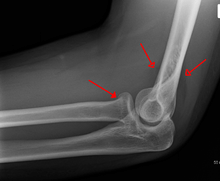
A subtle radial head fracture with associated positive
sail signSpecific fracture types of the radius include:
Gallery
| Position of radius (shown in red). |
| Radius, styloid process - anterior view |
| Radius, ulnar notch - posterior view |
| Radius, radial head - posterior view |
| Radius, radial head - anterior view |
| Radius l. dx. - ant. view |
| Radius l. dx. - post. view |
| Right human radius and ulna - post. view |
| Bones of left forearm - ant. view |
| Bones of left forearm - post. view |
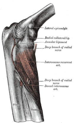
| Left elbow-joint, showing anterior and ulnar collateral ligaments |
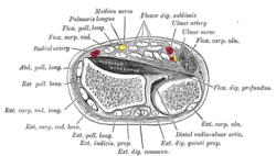
| Cross-section through middle of forearm |
| Transverse section across distal ends of radius and ulna |
| Muscles of upper limb. Cross section. |
| Wrist joint. Deep dissection. Posterior view. |
| Wrist joint. Deep dissection.Anterior, palmar, view. |
| Wrist joint. Deep dissection.Anterior, palmar, view. |
| Wrist joint. Deep dissection.Anterior, palmar, view. |
|
See also
- This article uses anatomical terminology; for an overview, see anatomical terminology.
References
This article incorporates text in the public domain from the 20th edition of Gray's Anatomy (1918)
- ↑ Marieb, E., R.N., Ph.D; Mallatt, J., Ph.D. & Wilhelm, P., Ph.D. (2008), Human Anatomy (5th ed.), San Francisco, CA: Pearson Benjamin Cummings, p. 188
- ↑ Clemente, Carmine D. (2007), Anatomy: A Regional Atlas of the Human Body (5th ed.), Philadelphia, PA: Lippincott Williams & Wilkins
- ↑ Romer, Alfred Sherwood; Parsons, Thomas S. (1977). The Vertebrate Body. Philadelphia, PA: Holt-Saunders International. p. 199. ISBN 0-03-910284-X.
- ↑ Essex Lopresti fracture at Wheeless' Textbook of Orthopaedics online
External links
|
|---|
| | Pectoral girdle, clavicle | |
|---|
| | Scapula | |
|---|
| | Humerus | |
|---|
| | Forearm | |
|---|
| | Hand | | Carpal bones | |
|---|
| | Metacarpal bones | |
|---|
| | Phalanges |
- proximal
- intermediate
- distal
|
|---|
|
|---|
| |
|---|
| | Description |
- Anatomy
- bones
- skull
- face
- neurocranium
- compound structures
- foramina
- upper extremity
- torso
- pelvis
- lower extremity
- Physiology
- Development
- Cells
|
|---|
| | Disease |
- Congenital
- Neoplasms and cancer
- Trauma
- Other
- Symptoms and signs
|
|---|
| | Treatment | |
|---|
|
|


.jpg)