Protein phosphorylation
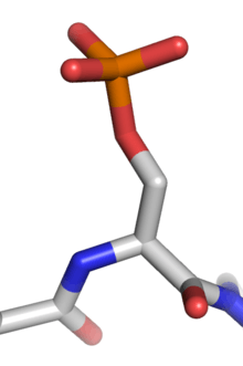
Protein phosphorylation is a post-translational modification of proteins in which an amino acid residue is phosphorylated by a protein kinase by the addition of a covalently bound phosphate group. Phosphorylation alters the structural conformation of a protein, causing it to become activated, deactivated, or modifying its function. The reverse reaction of phosphorylation is called dephosphorylation, and is catalyzed by protein phosphatases. Protein kinases and phosphatases work independently and in a balance to regulate the function of proteins. The amino acids most commonly phosphorylated are serine, threonine, and tyrosine in eukaryotes, and histidine in prokaryotes, which play important and well-characterized roles in signaling pathways and metabolism. However, many other amino acids can also be phosphorylated, including arginine, lysine, and cysteine.[1] Protein phosphorylation was first reported in 1906 by Phoebus Levene at the Rockefeller Institute for Medical Research with the discovery of phosphorylated vitellin.[2] However, it was nearly 50 years until the enzymatic phosphorylation of proteins by protein kinases was discovered.[3]
Functions of phosphorylation
Phosphorylation introduces a charged and hydrophobic element at the R group of the modified amino acid, changing a protein's structure by altering interactions with nearby amino acids. Some proteins such as p53 contain multiple phosphorylation sites, facilitating complex, multi-level regulation. Because of the ease with which proteins can be phosphorylated and dephosphorylated, this type of modification is a flexible mechanism for cells to respond to external signals and environmental conditions.[4]
The first example of protein regulation by phosphorylation was glycogen phosphorylase. Eddie Fisher and Ed Krebs described how phosphorylation of glycogen phosphorylase b converted it to the active glycogen phosphorylase a. It was soon discovered that glycogen synthase, another metabolic enzyme, is inactivated by phosphorylation.[5] Regulatory roles of phosphorylation include:
- Biological thermodynamics of energy-requiring reactions
- Phosphorylation of Na+/K+-ATPase during the transport of sodium (Na+) and potassium (K+) ions across the cell membrane in osmoregulation to maintain homeostasis of the body's water content.
- Mediates enzyme inhibition
- Phosphorylation of the enzyme GSK-3 by AKT (Protein kinase B) as part of the insulin signaling pathway.[6]
- Phosphorylation of src tyrosine kinase (pronounced "sarc") by C-terminal Src kinase (Csk) induces a conformational change in the enzyme, resulting in a fold in the structure, which masks its kinase domain, and is thus shut "off".[7]
- Important for protein-protein interaction via "recognition domains."
- Phosphorylation of the cytosolic components of NADPH oxidase, a large membrane-bound, multi-protein enzyme present in phagocytic cells, plays an important role in the regulation of protein-protein interactions in the enzyme.[8]
- Important in protein degradation.
- In the late 1990s, it was recognized that phosphorylation of some proteins causes them to be degraded by the ATP-dependent ubiquitin/proteasome pathway. These target proteins become substrates for particular E3 ubiquitin ligases only when they are phosphorylated.
Receptor Tyrosine Kinases
While tyrosine phosphorylation is found in relatively low abundance, it is well studied due to the ease of purification of phosphotyrosine using antibodies. Receptor tyrosine kinases are an important family of cell surface receptors involved in the transduction of extracellular signals such as hormones, growth factors, and cytokines. Binding of a ligand to a monomeric receptor tyrosine kinase stabilizes interactions between two monomers to form a dimer, after which the two bound receptors phosphorylate tyrosine residues in trans. Phosphorylation and activation of the receptor activates a signaling pathway through enzymatic activity and interactions with adaptor proteins.[9] Signaling through the epidermal growth factor receptor (EGFR), a receptor tyrosine kinase, is critical for the development of multiple organ systems including the skin, lung, heart, and brain. Excessive signaling through the EGFR pathway is found in many human cancers.[10]
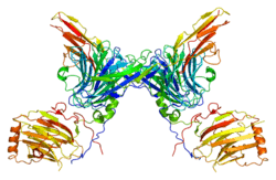
Cyclin-Dependent Kinases
Cyclin-dependent kinases (CDKs) are serine-threonine kinases which regulate progression through the eukaryotic cell cycle. CDKs are catalytically active only when bound to a regulatory cyclin. Animal cells contain at least nine distinct CDKs which bind to various cyclins with considerable specificity. CDK inhibitors (CKIs) block kinase activity in the cyclin-CDK complex to halt the cell cycle in G1 or in response to environmental signals or DNA damage. The activity of different CDKs activate cell signaling pathways and transcription factors that regulate key events in mitosis such as the G1/S phase transition. Earlier cyclin-CDK complexes provide the signal to activate subsequent cyclin-CDK complexes.[11]
Evolution
Protein phosphorylation is common among all clades of life, including all animals, plants, fungi, bacteria, and archaea. The origins of protein phosphorylation mechanisms are ancestral and have diverged greatly between different species. In eukaryotes, it is estimated that as many as 30% of all proteins may be phosphorylated, with tens of thousands of distinct phosphorylation sites.[12] Some phosphorylation sites appear to have evolved as conditional "off" switches, blocking the active site of an enzyme, such as in the prokaryotic metabolic enzyme isocitrate dehydrogenase. However, in the case of proteins that must be phosphorylated to be active, it is less clear how they could have emerged from non-phosphorylated ancestors. It has been shown that a subset of serine phosphosites are often replaced by acidic residues such as aspartate and glutamate between different species. These anionic residues can interact with cationic residues such as lysine and arginine to form salt bridges, stable non-covalent interactions that alter a protein's structure. These phosphosites often participate in salt bridges, suggesting that some phosphorylation sites evolved as conditional "on" switches for salt bridges, allowing these proteins to adopt an active conformation only in response to a specific signal.[13]
There are hundreds of known eukaryotic protein kinases, making them one of the largest gene families. Most phosphorylation is carried out by a single superfamily of protein kinases that share a conserved kinase domain. Protein phosphorylation is highly conserved in pathways central to cell survival, such as cell cycle progression relying on Cyclin-dependent kinases (CDKs), but individual phosphorylation sites are often flexible. Targets of CDK phosphorylation often have phosphosites in disordered segments, which are found in non-identical locations even in close species. Conversely, targets of CDK phosphorylation in structurally defined regions are more highly conserved. While CDK activity is critical for cell growth and survival in all eukaryotes, only very few phosphosites show strong conservation of their precise positions. Positioning is likely to be highly important for phosphates that allosterically regulate protein structure, but much more flexible for phosphates that interact with phosphopeptide-binding domains to recruit regulatory proteins.[14]
References
- ↑ Cieśla J, Frączyk T, Rode W (2011). "Phosphorylation of basic amino acid residues in proteins: important but easily missed". Acta Biochimica Polonica 58 (2): 137–147.
- ↑ Levene PA, Alsberg CL (1906). "The cleavage products of vitellin". J. Biol. Chem. 2 (1): 127–133.
- ↑ Burnett G, Kennedy EP (December 1954). "The enzymatic phosphorylation of proteins". J. Biol. Chem. 211 (2): 969–80. PMID 13221602.
- ↑ Johnson L N, Barford D. The effects of phosphorylation on the structure and function of proteins[J]. Annual review of biophysics and biomolecular structure, 1993, 22(1): 199-232.
- ↑ Johnson L N (2009). "The regulation of protein phosphorylation". Biochem Soc. Trans. 37 (2): 627–641. doi:10.1042/BST0370627.
- ↑ van Weeren PC, de Bruyn KM, de Vries-Smits AM, van Lint J, Burgering BM (May 1998). "Essential role for protein kinase B (PKB) in insulin-induced glycogen synthase kinase 3 inactivation. Characterization of dominant-negative mutant of PKB". J. Biol. Chem. 273 (21): 13150–6. doi:10.1074/jbc.273.21.13150. PMID 9582355.
- ↑ Cole PA, Shen K, Qiao Y, Wang D (October 2003). "Protein tyrosine kinases Src and Csk: a tail's tale". Curr Opin Chem Biol 7 (5): 580–5. doi:10.1016/j.cbpa.2003.08.009. PMID 14580561.
- ↑ Babior BM (March 1999). "NADPH oxidase: an update". Blood 93 (5): 1464–76. PMID 10029572.
- ↑ Lemmon MA, Schlessinger J (June 2010). "Cell signaling by receptor tyrosine kinases". Cell 141 (7): 1117–34. doi:10.1016/j.cell.2010.06.011. PMC 2914105. PMID 20602996.
- ↑ Cho HS, Leahy DJ; Leahy (2002). "Structure of the extracellular region of HER3 reveals an interdomain tether". Science 297 (5585): 1330–1333. Bibcode:2002Sci...297.1330C. doi:10.1126/science.1074611. PMID 12154198.
- ↑ Morgan, David O. (2007). The Cell Cycle: Principles of Control. London: New Science Press, 1st ed.
- ↑ Cohen P (2000). "The regulation of protein function by multisite phosphorylation – a 25 year update". Trends Biochem. Sci 25 (12): 596–601. doi:10.1016/S0968-0004(00)01712-6.
- ↑ Pearlman SM, Serber Z, Ferrell JE (2011). "A Mechanism for the Evolution of Phosphorylation Sites". Cell 147 (4): 934–946. doi:10.1016/j.cell.2011.08.052.
- ↑ Holt LJ, Tuch BB, Villén J, Johnson AD, Gygi SP, Morgan DO (2009). "Global Analysis of Cdk1 Substrate Phosphorylation Sites Provides Insights into Evolution". Science 325 (5948): 1682–1686. doi:10.1126/science.1172867.