Pineal gland
| Pineal gland | |
|---|---|
 Diagram of pituitary and pineal glands in the human brain | |
| Details | |
| Latin | glandula pinealis |
| Precursor | Neural Ectoderm, Roof of Diencephalon |
| posterior cerebral artery | |
| Identifiers | |
| Gray's | p.1277 |
| MeSH | Pineal+gland |
| NeuroLex ID | Pineal body |
| Dorlands /Elsevier | g_06/12392585 |
| TA | A11.2.00.001 |
| FMA | 62033 |
| Anatomical terms of neuroanatomy | |
The pineal gland, also known as the pineal body, conarium or epiphysis cerebri, is a small endocrine gland in the vertebrate brain. It produces melatonin, a serotonin derived hormone, which affects the modulation of sleep patterns in both seasonal and circadian rhythms.[1][2] Its shape resembles a tiny pine cone (hence its name), and it is located in the epithalamus, near the center of the brain, between the two hemispheres, tucked in a groove where the two halves of the thalamus join.
Nearly all vertebrate species possess a pineal gland. The most important exception is the hagfish, which is often thought of as the most primitive extant vertebrate.[3] Even in the hagfish, however, there may be a "pineal equivalent" structure in the dorsal diencephalon.[4] The lancelet Branchiostoma lanceolatum, the nearest existing relative to vertebrates, also lacks a recognizable pineal gland.[3] The lamprey (considered almost as primitive as the hagfish), however, does possess one.[3] A few more developed vertebrates, including the alligator, lack pineal glands because they have been lost over the course of evolution.[5]
The gland has been compared to the photoreceptive, so-called third parietal eye present in the epithalamus of some animal species, which is also called the pineal eye. René Descartes believed the pineal gland to be the "principal seat of the soul" and viewed it as the third eye.[6]
Structure
The pineal gland is the only midline brain structure that is unpaired (azygous). It takes its name from its pine-cone shape.[7] The gland is reddish-gray and about the size of a grain of rice (5–8 mm) in humans. The pineal gland, also called the pineal body, is part of the epithalamus, and lies between the laterally positioned thalamic bodies. It is located in the quadrigeminal cistern near to the corpora quadrigemina.[8] It is also located behind the third ventricle and is bathed in cerebrospinal fluid supplied through a small pineal recess of the third ventricle which projects into the stalk of the gland.[9]
Blood supply
Unlike most of the mammalian brain, the pineal gland is not isolated from the body by the blood–brain barrier system;[10] it has profuse blood flow, second only to the kidney.
Innervation
The pineal gland receives a sympathetic innervation from the superior cervical ganglion. A parasympathetic innervation from the pterygopalatine and otic ganglia is also present. Further, some nerve fibers penetrate into the pineal gland via the pineal stalk (central innervation). Also, neurons in the trigeminal ganglion innervate the gland with nerve fibers containing the neuropeptide PACAP.
Histology


The pineal body consists in humans of a lobular parenchyma of pinealocytes surrounded by connective tissue spaces. The gland's surface is covered by a pial capsule.
The pineal gland consists mainly of pinealocytes, but four other cell types have been identified. As it is quite cellular (in relation to the cortex and white matter), it may be mistaken for a neoplasm.[11]
| Cell type | Description |
|---|---|
| Pinealocytes | The pinealocytes consist of a cell body with 4–6 processes emerging. They produce and secrete melatonin. The pinealocytes can be stained by special silver impregnation methods. Their cytoplasm is lightly basophilic. With special stains, pinealocytes exhibit lengthy, branched cytoplasmic processes that extend to the connective septa and its blood vessels. |
| Interstitial cells | Interstitial cells are located between the pinealocytes. They have elongated nuclei and a cytoplasm that is stained darker than that of the pinealocytes. |
| Perivascular phagocyte | Many capillaries are present in the gland, and perivascular phagocytes are located close to these blood vessels. The perivascular phagocytes are antigen presenting cells. |
| pineal neurons | In higher vertebrates neurons are usually located in the pineal gland. However, this is not the case in rodents. |
| peptidergic neuron-like cells | In some species, neuronal-like peptidergic cells are present. These cells might have a paracrine regulatory function. |
In some parts of the brain and in particular the pineal gland, there are calcium structures, the number of which increases with age, called corpora arenacea (or "acervuli," or "brain sand"). Chemical analysis shows that they are composed of calcium phosphate, calcium carbonate, magnesium phosphate, and ammonium phosphate.[12] In 2002, deposits of the calcite form of calcium carbonate were described.[13] Calcium and phosphorus[14] deposits in the pineal gland have been linked with aging.
Development
The human pineal gland grows in size until about 1–2 years of age, remaining stable thereafter,[15][16] although its weight increases gradually from puberty onwards.[17][18] The abundant melatonin levels in children are believed to inhibit sexual development, and pineal tumors have been linked with precocious puberty. When puberty arrives, melatonin production is reduced.
Function
Melatonin is N-acetyl-5-methoxy-tryptamine, a derivative of the amino acid tryptophan, which also has other functions in the central nervous system. The production of melatonin by the pineal gland is stimulated by darkness and inhibited by light.[19][20] Photosensitive cells in the retina detect light and directly signal the suprachiasmatic nucleus (SCN), entraining its rhythm to the 24-hour cycle in nature. Fibers project from the SCN to the paraventricular nuclei (PVN), which relay the circadian signals to the spinal cord and out via the sympathetic system to superior cervical ganglia (SCG), and from there into the pineal gland.
The compound pinoline is also produced in the pineal gland; it is one of the beta-carbolines.[21]
Regulation of the pituitary gland
Studies on rodents suggest that the pineal gland influences the pituitary gland's secretion of the sex hormones follicle-stimulating hormone (FSH), and luteinizing hormone (LH). In a study by Motta, Fraschini, and Martini (1967), a pinealectomy was performed on rodents. No change in pituitary weight was observed, however there was an increase in the concentration of FSH and LH within the gland. In this same study, administration of melatonin did not return the concentrations of FSH to normal levels, suggesting that the pineal gland influences the pituitary glands secretion of FSH and LH through some other transmitting molecule.[22]
Drug metabolism
Studies on rodents suggest that the pineal gland may influence the actions of recreational drugs, such as cocaine,[23] and antidepressants, such as fluoxetine (Prozac),[24] and its hormone melatonin can protect against neurodegeneration.[25]
Clinical significance
Calcification
The pineal gland is often seen in skull X-rays, when it is calcified (in old age).[26]
Calcification of the pineal gland is typical in adults, and has been observed in children as young as 2. Calcification rates vary widely by country and correlate with an increase in age, with calcification occurring in an estimated 40% of Americans by their 17th year.[26] Calcification of the pineal gland is largely associated with corpora arenacea also known as "brain sand".
It seems that the internal secretions of the pineal gland inhibit the development of the reproductive glands, because, in cases where it is severely damaged in children, the result is accelerated development of the sexual organs and the skeleton.[27] In animals, the pineal gland appears to play a major role in sexual development, hibernation, metabolism, and seasonal breeding.[28]
Some recent studies show that the degree of pineal gland calcification is significantly higher in patients with Alzheimer's disease vs. other types of dementia.[29]
Pineal gland calcification may also contribute to the pathogenesis of Alzheimer's disease and may reflect an absence of crystallization inhibitors.[29]
Calcium, phosphorus,[14] and fluoride deposits in the pineal gland have been correlated with aging, showing that, as the brain ages, more deposits collect.[30]
Cancer
Tumors of the pineal gland are called pinealomas. These tumors are rare and 50% to 70% are germinomas that arise from sequestered embryonic germ cells. Histologically they are similar to testicular seminomas and ovarian dysgerminomas.
A pineal tumor can compress the superior colliculi and pretectal area of the dorsal midbrain, producing Parinaud's syndrome. Pineal tumors also can cause compression of the cerebral aqueduct, resulting in a noncommunicating hydrocephalus.
These neoplasms are divided into two categories, pineoblastomas and pineocytomas, based on their level of differentiation, which, in turn, correlates with their neoplastic aggressiveness. The clinical course of patients with pineocytomas is prolonged, averaging 7 years. The manifestations are the consequence of their pressure effects and consist of visual disturbances, headache, mental deterioration, and sometimes dementia-like behaviour.The position of these tumors makes them very difficult or impossible to remove surgically.
Other animals
Pinealocytes in many non-mammalian vertebrates have a strong resemblance to the photoreceptor cells of the eye. Some evolutionary biologists believe that the vertebrate pineal cells possess a common evolutionary ancestor with retinal cells.[31]
Pineal cytostructure seems to have evolutionary similarities to the retinal cells of chordates.[31] Modern birds and reptiles have been found to express the phototransducing pigment melanopsin in the pineal gland. Avian pineal glands are believed to act like the SCN in mammals.[32]
In some vertebrates, exposure to light can set off a chain reaction of enzymatic events within the pineal gland that regulate circadian rhythms.[33] Some early vertebrate fossil skulls have a pineal foramen (opening). This correlates with the physiology of the modern "living fossils," the lampreys and the tuatara, and some other vertebrates that have a parietal eye, which, in some of them, is photosensitive. The parietal eye represents evolution's earlier approach to photoreception.[34] The structures of the pineal eye in the tuatara are analogous to the cornea, lens, and retina, though the latter resembles that of an octopus rather than a vertebrate retina. The asymmetrical whole consists of the "eye" to the left and the pineal sac to the right. "In animals that have lost the parietal eye, including mammals, the pineal sac is retained and condensed into the form of the pineal gland."[34]
Fossils seldom preserve soft anatomy. The brain of the Russian Melovatka bird, about 90 million years old, is an exception, and it shows a larger-than-expected parietal eye and pineal gland.[35]
In humans and other mammals, the light signals necessary to set circadian rhythms are sent from the eye through the retinohypothalamic system to the suprachiasmatic nuclei (SCN) and the pineal gland.
Society and culture
René Descartes, dedicating much time to the study of the pineal gland, called it the "principal seat of the soul".[36] He believed it to be the point of connection between the intellect and the body.[37] Descartes attached significance to the gland because he believed it to be the only section of the brain to exist as a single part rather than one-half of a pair. He argued that, because a person can never have "more than one thought at a time," external stimuli must be united within the brain before being considered by the soul, and he considered the pineal gland to be situated in "the most suitable possible place for this purpose," located centrally in the brain and surrounded by branches of the carotid arteries.[36]
Baruch de Spinoza criticized Descartes' viewpoint for neither following from self-evident premises nor being "clearly and distinctly perceived" (Descartes having previously asserted that he could not draw conclusions of this sort), and questioned what Descartes meant by talking of "the union of the mind and the body."[38]
The notion of a "pineal-eye" is central to the philosophy of the French writer Georges Bataille, which is analyzed at length by literary scholar Denis Hollier in his study Against Architecture. In this work Hollier discusses how Bataille uses the concept of a "pineal-eye" as a reference to a blind-spot in Western rationality, and an organ of excess and delirium.[39] This conceptual device is explicit in his surrealist texts, The Jesuve and The Pineal Eye.[40]
Numerous spiritual philosophies contain the notion of an inner third eye that is related to the Ajna chakra and also to the pineal gland. These are attributed significance in mystical awakening or enlightenment, clairvoyant perception, and higher states of consciousness. This idea occurs historically in ancient central and eastern Asia, as well as in contemporary theories relating to yoga, theosophy, Hinduism and pagan religions, and New Age spiritual philosophies.
History
The secretory activity of the pineal gland is only partially understood. Its location deep in the brain suggested to philosophers throughout history that it possesses particular importance. This combination led to its being regarded as a "mystery" gland with mystical, metaphysical, and occult theories surrounding its perceived functions.
The pineal gland was originally believed to be a "vestigial remnant" of a larger organ. In 1917, it was known that extract of cow pineals lightened frog skin. Dermatology professor Aaron B. Lerner and colleagues at Yale University, hoping that a substance from the pineal might be useful in treating skin diseases, isolated and named the hormone melatonin in 1958.[41] The substance did not prove to be helpful as intended, but its discovery helped solve several mysteries such as why removing the rat's pineal accelerated ovary growth, why keeping rats in constant light decreased the weight of their pineals, and why pinealectomy and constant light affect ovary growth to an equal extent; this knowledge gave a boost to the then new field of chronobiology.[42]
See also
Additional images
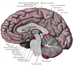
The pineal body is labeled in these images.
-

Mesal aspect of a brain sectioned in the median sagittal plane.
-
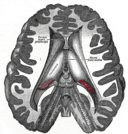
Dissection showing the ventricles of the brain.
-

Hind- and mid-brains; antero-lateral view.
-
Median sagittal section of brain.
-

Pineal gland
-

Brainstem. Posterior view.
References
- ↑ Macchi M, Bruce J (2004). "Human pineal physiology and functional significance of melatonin". Front Neuroendocrinol 25 (3–4): 177–95. doi:10.1016/j.yfrne.2004.08.001. PMID 15589268.
- ↑ Arendt J, Skene DJ (2005). "Melatonin as a chronobiotic". Sleep Med Rev 9 (1): 25–39. doi:10.1016/j.smrv.2004.05.002. PMID 15649736.
Exogenous melatonin has acute sleepiness-inducing and temperature-lowering effects during 'biological daytime', and when suitably timed (it is most effective around dusk and dawn) it will shift the phase of the human circadian clock (sleep, endogenous melatonin, core body temperature, cortisol) to earlier (advance phase shift) or later (delay phase shift) times.
- ↑ 3.0 3.1 3.2 Vernadakis AJ, Bemis WE, Bittman EL (April 1998). "Localization and partial characterization of melatonin receptors in amphioxus, hagfish, lamprey, and skate". Gen. Comp. Endocrinol. 110 (1): 67–78. doi:10.1006/gcen.1997.7042. PMID 9514841.
- ↑ Ooka-Souda S, Kadota T, Kabasawa H (December 1993). "The preoptic nucleus: the probable location of the circadian pacemaker of the hagfish, Eptatretus burgeri". Neurosci. Lett. 164 (1–2): 33–6. doi:10.1016/0304-3940(93)90850-K. PMID 8152610.
- ↑ Erlich SS, Apuzzo ML (September 1985). "The pineal gland: anatomy, physiology, and clinical significance". J. Neurosurg. 63 (3): 321–41. doi:10.3171/jns.1985.63.3.0321. PMID 2862230.
- ↑ Eakin, R. M (1973). The Third Eye. Berkeley: University of California Press.
- ↑ Bowen, R. "The Pineal Gland and Melatonin". Retrieved 14 October 2011.
- ↑ http://www.ajnr.org/content/19/9/1631.full.pdf
- ↑ Dorland's. Illustrated Medical Dictionary. Elsevier Saunders. p. 1607. ISBN 978-1-4160-6257-8.
- ↑ Pritchard, Thomas C.; Alloway, Kevin Douglas (1999). Medical Neuroscience (GOOGLE BOOKS PREVIEW). Hayes Barton Press. pp. 76–77. ISBN 1-889325-29-5. Retrieved 2009-02-08.
- ↑ Kleinschmidt-DeMasters BK, Prayson RA (November 2006). "An algorithmic approach to the brain biopsy—part I". Arch. Pathol. Lab. Med. 130 (11): 1630–8. doi:10.1043/1543-2165(2006)130[1630:AAATTB]2.0.CO;2. PMID 17076524.
- ↑ Bocchi G, Valdre G (1993). "Physical, chemical, and mineralogical characterization of carbonate-hydroxyapatite concretions of the human pineal gland". J Inorg Biochem 49 (3): 209–20. doi:10.1016/0162-0134(93)80006-U. PMID 8381851.
- ↑ Baconnier S, Lang S, Polomska M, Hilczer B, Berkovic G, Meshulam G (2002). "Calcite microcrystals in the pineal gland of the human brain: first physical and chemical studies". Bioelectromagnetics 23 (7): 488–95. doi:10.1002/bem.10053. PMID 12224052.
- ↑ 14.0 14.1 "IngentaConnect High Accumulation of Calcium and Phosphorus in the Pineal Bodies". Ingentaconnect.com. 2006-06-16. Retrieved 2009-07-06.
- ↑ Lack of pineal growth during childhood. Schmidt F, Penka B, Trauner M, Reinsperger L, Ranner G, Ebner F, Waldhauser F. J Clin Endocrinol Metab. 1995 Apr;80(4):1221–5.
- ↑ Development of the pineal gland: measurement with MR. Sumida M, Barkovich AJ, Newton TH. AJNR Am J Neuroradiol. 1996 Feb;17(2):233–6.
- ↑ Tapp E, Huxley M. The weight and degree of calcification of the pineal gland. J Pathol 1971;105:31–39
- ↑ Tapp E, Huxley M. The histological appearance of the human pineal gland from puberty to old age. J Pathol 1972;108:137–144
- ↑ Axelrod J (1970). "The pineal gland". Endeavour 29 (108): 144–8. PMID 4195878.
- ↑ Lowrey, Phillip L., and Joseph S. Takahashi. "Genetics of the mammalian circadian system: photic entrainment, circadian pacemaker mechanisms, and posttranslational regulation." Annual review of genetics 34.1 (2000): 533-562.
- ↑ Callaway, James C.; Gyntber, Jukka; Poso, Antti; Airaksinen, Mauno M.; Vepsäläinen, Jouko (1994). "The pictet-spengler reaction and biogenic tryptamines: Formation of tetrahydro-β-carbolines at physiologicalpH". Journal of Heterocyclic Chemistry 31 (2): 431. doi:10.1002/jhet.5570310231.
- ↑ Motta, Marina; Fraschini, F.; Martini, L. (1967). "Endocrine Effects of Pineal Gland and of Melatonin.". Exp Biol Med (Maywood) 126 (2): 431–435. doi:10.3181/00379727-126-32468.
- ↑ Uz T, Akhisaroglu M, Ahmed R, Manev H (2003). "The pineal gland is critical for circadian Period1 expression in the striatum and for circadian cocaine sensitization in mice". Neuropsychopharmacology 28 (12): 2117–23. doi:10.1038/sj.npp.1300254. PMID 12865893.
- ↑ Uz T, Dimitrijevic N, Akhisaroglu M, Imbesi M, Kurtuncu M, Manev H (2004). "The pineal gland and anxiogenic-like action of fluoxetine in mice". Neuroreport 15 (4): 691–4. doi:10.1097/00001756-200403220-00023. PMID 15094477.
- ↑ Manev H, Uz T, Kharlamov A, Joo J (1996). "Increased brain damage after stroke or excitotoxic seizures in melatonin-deficient rats". FASEB J 10 (13): 1546–51. PMID 8940301.
- ↑ 26.0 26.1 Zimmerman, Robert A. "Age-Related Incidence of Pineal Calcification Detected by Computed Tomography" (PDF). Radiological Society of North America. Retrieved 21 June 2012.
- ↑ "The Pineal Body". Human Anatomy (Gray's Anatomy). Retrieved 2011-09-07.
- ↑ Strassman
- ↑ 29.0 29.1 Mahlberg, R.; Walther, S.; Kalus, P.; Bohner, G.; Haedel, S.; Reischies, F. M.; Kühl, K. P.; Hellweg, R.; Kunz, D. (2008). "Pineal calcification in Alzheimer's disease: An in vivo study using computed tomography". Neurobiology of Aging 29 (2): 203–209. doi:10.1016/j.neurobiolaging.2006.10.003. PMID 17097768.
- ↑ Luke, Jennifer (March–April 2001). "Fluoride Deposition in the Aged Human Pineal Gland". Caries Res 2001 (35): 125–128. doi:10.1159/000047443. PMID 11275672.
- ↑ Natesan A, Geetha L, Zatz M (2002). "Rhythm and soul in the avian pineal". Cell Tissue Res 309 (1): 35–45. doi:10.1007/s00441-002-0571-6. PMID 12111535.
- ↑ Moore RY, Heller A, Wurtman RJ, Axelrod J (January 1967). "Visual pathway mediating pineal response to environmental light". Science 155 (759): 220–3. doi:10.1126/science.155.3759.220. PMID 6015532.
- ↑ 34.0 34.1 Schwab, I.R.; O'Connor, G.R. (March 2005). "The lonely eye". British Journal of Ophthalmology 89 (3): 256. doi:10.1136/bjo.2004.059105. PMC 1772576. PMID 15751188.
- ↑ Kurochkin, Evgeny N.; Gareth J. Dyke; Sergei V. Saveliev; Evgeny M. Pervushov; Evgeny V. Popov (June 2007). "A fossil brain from the Cretaceous of European Russia and avian sensory evolution". Biology Letters (The Royal Society) 3 (3): 309–313. doi:10.1098/rsbl.2006.0617. PMC 2390680. PMID 17426009.
- ↑ 36.0 36.1 Descartes and the Pineal Gland (Stanford Encyclopedia of Philosophy)
- ↑ Descartes R. "The Passions of the Soul" excerpted from "Philosophy of the Mind," Chalmers, D. New York: Oxford University Press, Inc.; 2002. ISBN 978-0-19-514581-6
- ↑ http://en.wikisource.org/wiki/Ethics_%28Spinoza%29/Part_5
- ↑ Hollier, D, Against Architecture: The Writings of Georges Bataille, trans. Betsy Wing, MIT, 1989.
- ↑ Bataille, G, Visions of Excess: Selected Writings, 1927–1939 (Theory and History of Literature, Vol 14), trans. Allan Stoekl et al., Manchester University Press, 1985
- ↑ Lerner AB, Case JD, Takahashi Y (1960). "Isolation of melatonin and 5-methoxyindole-3-acetic acid from bovine pineal glands". J Biol Chem 235: 1992–7. PMID 14415935.
- ↑ Coates, Paul M. (2005). Encyclopedia of Dietary Supplements. Marc R. Blackman, Gordon M. Cragg, Mark Levine, Joel Moss, Jeffrey D. White. CRC Press. p. 457. ISBN 0-8247-5504-9. Retrieved 2009-03-31.
External links
| Look up pineal gland in Wiktionary, the free dictionary. |
| Wikimedia Commons has media related to Pineal gland. |
- Stained brain slice images which include the "pineal%20gland" at the BrainMaps project
- hier-280 at NeuroNames
- Histology at BU: Endocrine System: pineal gland (illustration)
- Anatomy Atlases, Microscopic atlas: Pineal gland
- MedPix: Images of Pineal region
| ||||||||||||||||||||||||||||||||||||||||||||||||||||||||||||||
| ||||||||||||||||||||||||||||||||||||||||||||||||||||||||||||||||||||||||||||
| ||||||||||||||||||||||||||||||||||||||||||||||||||||||||||||||||