Peranema
| Peranema | |
|---|---|
 | |
| Scientific classification | |
| Domain: | Eukaryota |
| Kingdom: | Protista |
| Phylum: | Euglenozoa |
| Class: | Euglenoidea |
| Order: | Euglenales |
| Family: | Peranemaceae |
| Genus: | Peranema Dujardin, 1841 |
| Species | |
|
See text | |
Peranema is a genus of free-living flagellate protists, with more than 20 accepted species, varying in size between 8 and 200 micrometers.[1] They are found in freshwater lakes, ponds and ditches, and are often abundant at the bottom of stagnant pools rich in decaying organic material.[2] Although they belong to the class Euglenoidea, and are morphologically similar to the green Euglena, Peranema have no chloroplasts, and cannot feed by autotrophy. Instead, they capture live prey, such as yeast, bacteria and other flagellates, consuming them with the help of a rigid feeding apparatus called a "rod-organ." Unlike the green Euglenids, they lack both an eyespot (stigma), and the paraflagellar body (photoreceptor) that is normally coupled with that organelle.[3] However, while Peranema lack a localized photoreceptor, they do possess the light-sensitive protein rhodopsin, and respond to changes in light with a characteristic "curling behaviour."[4]

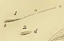
The earliest record of a Peranema is in O.F. Müller's Animalcula Infusoria of 1786, which describes an "elongated linear" creature, "stretched out at the front." Müller named it Vibrio strictus, placing it among the "long-necked" infusoria, along with Lacrymaria olor and Dileptus.[5] The species Peranema trichophorum was seen and described in 1838 by C.G. Ehrenberg, who, like Müller before him, took the flagellum for a necklike extension of the body, and placed it in the ciliate genus Trachelius.[6] Peranema was correctly identified as a flagellate by Félix Dujardin, who created the genus in 1842, giving it the name Pyronema, for its pyriform (pear-shaped) body. However, because that name had already been applied to a genus of fungi, he amended the genus to Peranema, formed from the Greek πέρα (a leather purse or sack) and νήμα (a thread).[7] Unfortunately, this name had also been claimed earlier, for a genus of ferns first collected in Nepal. As a result, botanists, following the International Code of Botanical Nomenclature, customarily refer to the protist Peranema as Pseudoperanema; whereas protozoologists, following the International Code of Zoological Nomenclature, have continued to call the genus by the name Dujardin gave it.[8]
Appearance and characteristics

Peranema's basic anatomy is that of a typical Euglenid. The cell is spindle or cigar-shaped, somewhat pointed at the anterior end. It has a pellicle with finely-ridged microtubules (a structure often referred to as "pellicle strips") arranged in a helical fashion around the body. On this type of pellicle, which is shared by many Euglenids, the spiraling microtubular strips are able to slide past one another, giving the organism an extremely plastic and changeable body shape. This permits a type of squirming motility, sometimes referred to as "Euglenoid movement" or "metaboly".[9] When it is not swimming, Peranema can creep along by metaboly, progressing with wavelike contractions of the body, reminiscent of peristalsis.[10][11]
At the anterior of the cell, there is a narrow aperture, opening into a flask-shaped "reservoir", from which the organism's two flagella emerge. At the bottom of this reservoir lie the basal bodies (centrioles) to which the flagella are attached.[12] One flagellum is relatively long and conspicuous, and when the Peranema is swimming it is held stiffly in front. At the tip of the flagellum, a short segment beats and flails in a rhythmic manner, causing the Peranema to move forward through the water with a calm, gliding motion. Peranama usually swims belly-down, without rotating.[13]
The second flagellum is difficult to see with bright field microscopy, and was entirely overlooked by early observers. It emerges from the same reservoir as the larger propulsive flagellum, but turns toward the posterior. It does not float freely, like the trailing flagella of Dinema and Entosiphon, but adheres to the outside of the cell membrane, in a groove along its ventral surface.[14]
Next to the reservoir, lies Peranema's highly developed feeding apparatus, a cytostomal sac supported on one side by a pair of rigid rods, fused together at the forward end. The use of this "rod-organ" in feeding has attracted considerable scholarly interest. Some early researchers speculated that it might assist Peranema in tearing up and consuming its food; while others held that it was actually a tubular construction, serving as a cytopharynx.[15] In 1950, Y. T. Chen accurately identified it as a structure separate from the reservoir, which could be used by Peranama to cut and pierce its prey.[16] Brenda Nisbet questioned this, on the grounds that, when examined closely with an electron microscope, the rod-organ is blunt, and therefore an improbable instrument for either cutting or piercing. Since the rod-organ had been seen to move back and forth during feeding, Nisbet argued that its primary function is to create suction, drawing prey into the cytostome.[12]
In 1997, Richard Triemer returned to the subject, to confirm Chen's opinion that Peranema has a dual feeding technique. It can swallow prey whole, pulling large flagellates through the cytostome, in a manner similar to that proposed by Brenda Nisbet. However, it can also choose a more elaborate style of attack. Sometimes, it will press its cytostome against its prey, and then move the rod-organ up and down, using a rasping motion to chew a hole in its victim's cell membrane. After consuming some of the protoplasm, the Peranema may then insert its large flagellum into the hole, using it to churn up the contents of the cell so that they may be more easily sucked out. This continues until nothing is left of the prey but the tattered remnants of its pellicle.[17]
Phylogeny and classification

When Dujardin created the genus Peranema in 1841, he was unable to detect the second flagellum and classified it with other ostensibly uniflagellate "Eugléniens," Astasia and Euglena. In 1881 Georg Klebs drew a taxonomical distinction between colorless uniflagellates that live by phagotrophy (Peranema and Astasia) and the green uniflagellates that photosynthesize (Euglena).[18] This distinction was generally abandoned after the publication, in 1952, of a major revision of the Euglenoids.[19] In 1997, a combined morphological and molecular analysis of certain Euglenoids identified Peranama trichophorum, Euglena gracilis and Khawkinea quartana as a distinct monophyletic lineage, with P. trichophorum basal to the other two species.[20]
Video Gallery
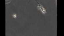
 Video of Peranema sp. in phase contrast microscopy, from the YouTube channel of microuruguay. |
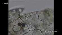
 Video of Peranema sp., showing the ingestion apparatus, from the YouTube channel of microuruguay. |
Species
- Peranema asperum, Playfair
- Peranema asperum var. ''rectangulare, Playfair
- Peranema cryptocercum, (Skuja) Popova
- Peranema cuneatum, Playfair
- Peranema curvicauda, Skuja
- Peranema deflexum, Skuja
- Peranema dolichonema
- Peranema furcatum, Skvortzov
- Peranema fusiforme, Larsen, 1987
- Peranema glabrum, Van Ove
- Peranema globulosa, F. Dujardin
- Peranema granuliferum, Penard
- Peranema hyalinum, Christen
- Peranema inflexum, Skuja
- Peranema kupfferi, Skuja
- Peranema limax, Christen
- Peranema nigrum, Christen
- Peranema ovale, Lackey
- Peranema macrostoma, Ekebom, Patterson & Vors
- Peranema macromastix, Conrad
- Peranema pleururum, Skuja
- Peranema sacculus, Christen
- Peranema trichophorum, (Ehrenberg) Stein
- Peranema truncatum, Skvortzov
Further Reading
Hassett, Charles (July 1944). "Photo-dynamic Action in the Flagellate Peranema Trichophorum with Special Reference to Motor Response to Light". Chicago Journals 17 (3): 270–278. Retrieved 13 February 2015.
References
- ↑ "Encyclopedia of Life".
- ↑ Mast, SO (Mar–Apr 1912). "The reactions of the flagellate Peranema". Journal of Animal Behaviour 2 (2): 91–97. doi:10.1037/h0072097.
- ↑ Brown, Virginius E. (1930). "The Cytology and Binary Fission of Peranema" (PDF). Quarterly Journal of Microscopical Science. 2 (73): 6.
- ↑ Saranak, Jureepan; Foster, Kenneth W. (2005). "Photoreceptor for Curling Behavior in Peranema trichophorum and Evolution of Eukaryotic Rhodopsins". Eukaryotic Cell 4 (10): 1605–1612. doi:10.1128/EC.4.10.1605-1612.2005.
- ↑ Müller, OF (1786). Animalcula Infusoria: Fluvia, Tilia et Marina. Hauniae, Typis N. Mölleri. p. 71.
- ↑ Saville-Kent, William (1882). A Manual of the Infusoria. Vol I. London: David Bogue.
- ↑ Dujardin, Felix (1841). Histoire Naturelle des Zoophytes. Infusoires. Vol. I. Paris: Roret.
- ↑ Patterson, David J.; Larsen, Jacob (January 1992). "A Perspective on Protistan Nomenclature". Journal of Eukaryotic Microbiology 39 (1): 126. doi:10.1111/j.1550-7408.1992.tb01292.x.
- ↑ Suzaki, T; Williamson, RE (1985). "Euglenoid Movement in Euglena fusca". Protoplasma 124 (1–2): 137–146. doi:10.1007/BF01279733.
- ↑ Chang, Shih L. (January 1966). "Observations on the Organelles, Movement, and Feeding of Peranema trichophorum (Ehrb.) Stein". Transactions of the American Microscopical Society 85 (1): 29–45. doi:10.2307/3224773. JSTOR 3224773.
- ↑ Chen, Y. T. (September 1950). "Investigations of the Biology of Peranema trichophorum (Euglenineae)" (PDF). Quarterly Journal of Microscopical Science. 3 (91): 302.
- ↑ 12.0 12.1 Nisbet, Brenda (February 1974). "An Ultrastructural Study of the Feeding Apparatus of Peranema trichophorum". Journal of Eukaryotic Microbiology 21 (1): 39–48. doi:10.1111/j.1550-7408.1974.tb03614.x.
- ↑ Jahn, Theodore Louis (1949). How to Know the Protozoa. Dubuque, Iowa: Wm. C. Brown. p. 72. ISBN 0-697-04829-2.
- ↑ Patterson, D.J. (1996) [1992]. Free-Living Freshwater Protozoa: A Colour Guide. Washington: Manson. p. 51. ISBN 1-874545-40-5.
- ↑ Pitelka, Dorothy Riggs (May 1945). "Morphology and taxonomy of flagellates of the genus Peranema Dujardin". Journal of Morphology 76 (3): 179–192. doi:10.1002/jmor.1050760304.
- ↑ Chen, Y. T. (September 1950). "Investigations of the Biology of Peranema trichophorum (Euglenineae)" (PDF). Quarterly Journal of Microscopical Science. 3 (91): 279–302.
- ↑ Triemer, Richard E. (August 1997). "FEEDING IN PERANEMA TRICHOPHORUM REVISITED (EUGLENOPHYTA)". Journal of Phycology 33 (4): 649–654. doi:10.1111/j.0022-3646.1997.00649.x.
- ↑ Pringsheim, E.G. (1948). "Taxonomic Problems in the Euglenineae". Biological Reviews 23 (1): 46–61. doi:10.1111/j.1469-185X.1948.tb00456.x. PMID 18901101.
- ↑ Linton, Eric W. et al. (March 1999). "A Molecular Study of Euglenoid Phylogeny using Small Subunit rDNA". Journal of Eukaryotic Microbiology 46 (2): 217–223. doi:10.1111/j.1550-7408.1999.tb04606.x. PMID 10361741.
- ↑ Montegut-Felkner, Anne E.; Triemer, Richard (June 1997). "PHYLOGENETIC RELATIONSHIPS OF SELECTED EUGLENOID GENERA BASED ON MORPHOLOGICAL AND MOLECULAR DATA". Journal of Phycology 33 (3): 512. doi:10.1111/j.0022-3646.1997.00512.x.
| ||||||||||||||||||||||||||||||||||||||||||||||||||||||||||||||||||||||||||||||||||||||||||
| ||||||||||||||||||||||||||||||||||||||||||||||||||||||||||||||||||||||||||||||||