Parathyroid hormone


Parathyroid hormone (PTH), parathormone or parathyrin, is secreted by the chief cells of the parathyroid glands as a polypeptide containing 84 amino acids. It acts to increase the concentration of calcium (Ca2+) in the blood, whereas calcitonin (a hormone produced by the parafollicular cells (C cells) of the thyroid gland) acts to decrease calcium concentration. PTH acts to increase the concentration of calcium in the blood by acting upon the parathyroid hormone 1 receptor (high levels in bone and kidney) and the parathyroid hormone 2 receptor (high levels in the central nervous system, pancreas, testis, and placenta).[1] PTH half-life is approximately 4 minutes.[2] It has a molecular mass of 9.4 kDa.[3]
Structure
hPTH-(1-34) crystallizes as a slightly bent, long helical dimer. Analysis reveals that the extended helical conformation of hPTH-(1-34) is the likely bioactive conformation.[4] The N-terminal fragment 1-34 of parathyroid hormone (PTH) has been crystallized and the structure has been refined to 0.9 Å resolution.
 helical dimer structure of hPTH-(1-34)[5] |
Function
Regulation of serum calcium
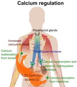
Parathyroid hormone regulates serum calcium through its effects on the following tissues:[6]
| Region | Effect |
| bone | It enhances the release of calcium from the large reservoir contained in the bones.[7] Bone resorption is the normal destruction of bone by osteoclasts, which are indirectly stimulated by PTH. Stimulation is indirect since osteoclasts do not have a receptor for PTH; rather, PTH binds to osteoblasts, the cells responsible for creating bone. Binding stimulates osteoblasts to increase their expression of RANKL and inhibits their expression of Osteoprotegerin (OPG). OPG binds to RANKL and blocks it from interacting with RANK, a receptor for RANKL. The binding of RANKL to RANK (facilitated by the decreased amount of OPG available for binding the excess RANKL) stimulates these osteoclast precursors to fuse, forming new osteoclasts, which ultimately enhances bone resorption. |
| kidney | It enhances active reabsorption of calcium and magnesium from distal tubules and the thick ascending limb. As bone is degraded, both calcium and phosphate are released. It also decreases the reabsorption of phosphate, with a net loss in plasma phosphate concentration. When the calcium:phosphate ratio increases, more calcium is free in the circulation.[8] |
| intestine via kidney | It enhances the absorption of calcium in the intestine by increasing the production of activated vitamin D. Vitamin D activation occurs in the kidney. PTH up-regulates 25-hydroxyvitamin D3 1-alpha-hydroxylase, the enzyme responsible for 1-alpha hydroxylation of 25-hydroxy vitamin D, converting vitamin D to its active form (1,25-dihydroxy vitamin D). This activated form of vitamin D increases the absorption of calcium (as Ca2+ ions) by the intestine via calbindin. |
PTH was one of the first hormones to be shown to use the G-protein, adenylyl cyclase second messenger system.
Normal total plasma calcium level ranges from 8.5 to 10.2 mg/dL (2.12 mmol/L to 2.55 mmol/L).[10]
Regulation of serum phosphate
PTH reduces the reabsorption of phosphate from the proximal tubule of the kidney,[11] which means more phosphate is excreted through the urine.
However, PTH enhances the uptake of phosphate from the intestine and bones into the blood. In the bone, slightly more calcium than phosphate is released from the breakdown of bone. In the intestines, absorption of both calcium and phosphate is mediated by an increase in activated vitamin D. The absorption of phosphate is not as dependent on vitamin D as is that of calcium. The end result of PTH release is a small net drop in the serum concentration of phosphate.
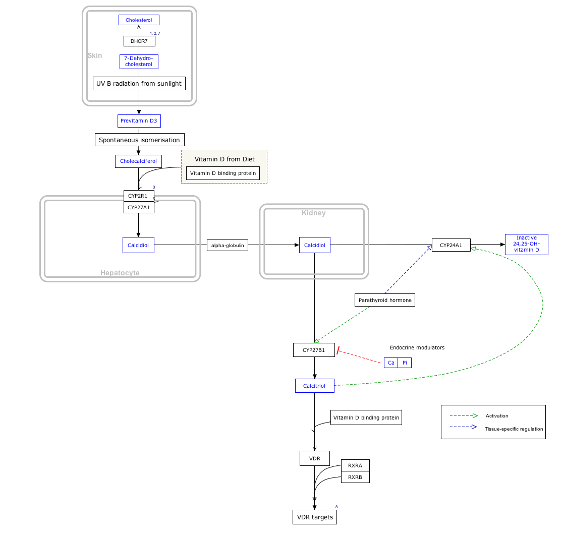
Vitamin D synthesis
PTH increases the activity of 1-α-hydroxylase enzyme, which converts 25-hydroxycholecalciferol to 1,25-dihydroxycholecalciferol, the active form of vitamin D.
Interactive pathway map
Click on genes, proteins and metabolites below to link to respective articles. [§ 1]
Vitamin D Synthesis Pathway edit
- ↑ The interactive pathway map can be edited at WikiPathways: "VitaminDSynthesis_WP1531".
Regulation of PTH secretion
Secretion of parathyroid hormone is controlled chiefly by serum [Ca2+] through negative feedback. Calcium-sensing receptors located on parathyroid cells are activated when [Ca2+] is elevated. The G-protein coupled calcium receptors bind extracellular calcium and may be found on the surface on a wide variety of cells distributed in the brain, heart, skin, stomach, C cells, and other tissues. In the parathyroid gland, high concentrations of extracellular calcium result in activation of the Gq G-protein coupled cascade through the action of phospholipase C. This hydrolyzes phosphatidylinositol 4,5-bisphosphate (PIP2) to liberate intracellular messengers IP3 and diacylglycerol (DAG). Ultimately, these two messengers result in a release of calcium from intracellular stores and a subsequent flux of extracellular calcium into the cytoplasmic space. The effect of this signaling of high extracellular calcium results in an intracellular calcium concentration that inhibits the secretion of preformed PTH from storage granules in the parathyroid gland. In contrast to the mechanism that most secretory cells use, calcium inhibits vesicle fusion and release of PTH.
In the parathyroids, magnesium serves this role in stimulus-secretion coupling. A mild decrease in serum magnesium levels stimulates the resorptive activity PTH has on the kidneys. Severe Hypomagnesemia inhibits PTH secretion and also causes resistance to PTH, leading to a form of hypoparathyroidism that is reversible.[12]
Stimulators
- Decreased serum [Ca2+].
- Mild decreases in serum [Mg2+].
- An increase in serum phosphate (increased phosphate causes it to complex with serum calcium, forming calcium phosphate, which reduces stimulation of Ca-sensitive receptors (CaSr) that do not sense calcium phosphate, triggering an increase in PTH).
Inhibitors
- Increased serum [Ca2+].
- Severe decreases in serum [Mg2+], which also produces symptoms of hypoparathyroidism (such as hypocalcemia).[13]
- Calcitriol
Clinical significance
- Hyperparathyroidism, the presence of excessive amounts of parathyroid hormone in the blood, occurs in two very distinct sets of circumstances. Primary hyperparathyroidism is due to autonomous, abnormal hypersecretion of PTH from the parathyroid gland, while secondary hyperparathyroidism is an appropriately high PTH level seen as a physiological response to hypocalcaemia.
- A low level of PTH in the blood is known as hypoparathyroidism and is most commonly due to damage to or removal of parathyroid glands during thyroid surgery.
- There are a number of rare but well-described genetic conditions affecting parathyroid hormone metabolism, including pseudohypoparathyroidism, familial hypocalciuric hypercalcaemia, and autosomal dominant hypercalciuric hypocalcaemia.
- In osteoporotic women, administration of an exogenous parathyroid hormone analogue (teriparatide, by daily injection) superimposed on estrogen therapy produced increases in bone mass and reduced vertebral and nonvertebral fractures by 45 to 65%.[14]
Measurement
PTH can be measured in the blood in several different forms: intact PTH; N-terminal PTH; mid-molecule PTH, and C-terminal PTH, and different tests are used in different clinical situations.
The average PTH level is 8–51 pg/ml.[15]
See also
- Calcium metabolism
- Disorders of calcium metabolism
- Parathyroid hormone family
- Parathyroid hormone-related protein
References
- ↑ Physiology at MCG 5/5ch6/s5ch6_11
- ↑ Bieglmayer C, Prager G, Niederle B (October 2002). "Kinetic analyses of parathyroid hormone clearance as measured by three rapid immunoassays during parathyroidectomy". Clin. Chem. 48 (10): 1731–8. PMID 12324490.
- ↑ Prahalad AK, Hickey RJ, Huang J et al. (June 2006). "Serum proteome profiles identifies parathyroid hormone physiologic response". Proteomics 6 (12): 3482–93. doi:10.1002/pmic.200500929. PMID 16705755.
- ↑ Jin L, Briggs SL, Chandrasekhar S, Chirgadze NY, Clawson DK, Schevitz RW, Smiley DL, Tashjian AH, Zhang F (September 2000). "Crystal structure of human parathyroid hormone 1-34 at 0.9-A resolution". J. Biol. Chem. 275 (35): 27238–44. doi:10.1074/jbc.M001134200. PMID 10837469.
- ↑ PDB 1ETE; Savvides SN, Boone T, Andrew Karplus P (June 2000). "Flt3 ligand structure and unexpected commonalities of helical bundles and cystine knots". Nature Structural & Molecular Biology 7 (6): 486–491. doi:10.1038/75896. PMID 10881197.; rendered via PyMOL.
- ↑ Coetzee M, Kruger MC (May 2004). "Osteoprotegerin-receptor activator of nuclear factor-kappaB ligand ratio: a new approach to osteoporosis treatment?". South. Med. J. 97 (5): 506–11. doi:10.1097/00007611-200405000-00018. PMID 15180028.
- ↑ Poole K, Reeve J (2005). "Parathyroid hormone - a bone anabolic and catabolic agent". Curr Opin Pharmacol 5 (6): 612–7. doi:10.1016/j.coph.2005.07.004. PMID 16181808.
- ↑ http://sprojects.mmi.mcgill.ca/nephrology/presentation/presentation5.htm
- ↑ Page 1094 (The Parathyroid Glands and Vitamin D) in: Walter F., PhD. Boron (2003). Medical Physiology: A Cellular And Molecular Approaoch. Elsevier/Saunders. p. 1300. ISBN 1-4160-2328-3.
- ↑ Zieve, MD, MHA, David. "MedlinePlus Medical Encyclopedia: Serum calcium". National Library of Medicine, National Institutes of Health. Retrieved 2009-02-01.
- ↑ Gardner, David; Shoback, Dolores (2011). Greenspan's Basic & Clinical Endocrinology (9th ed.). McGraw Hill. p. 232. ISBN 978-0071622431.
- ↑ Agus ZS (July 1999). "Hypomagnesemia". J. Am. Soc. Nephrol. 10 (7): 1616–22. PMID 10405219.
- ↑ Costanzo, Linda S. (2007). BRS Physiology. Lippincott, Williams, & Wilkins. p. 260. ISBN 978-0-7817-7311-9.
- ↑ Neer RM, Arnaud CD, Zanchetta JR, Prince R, Gaich GA, Reginster JY, Hodsman AB, Eriksen EF, Ish-Shalom S, Genant HK, Wang O, Mitlak BH (May 2001). "Effect of parathyroid hormone (1-34) on fractures and bone mineral density in postmenopausal women with osteoporosis". N. Engl. J. Med. 344 (19): 1434–41. doi:10.1056/NEJM200105103441904. PMID 11346808.
- ↑ Longo et al. Harrison's Principles of Internal Medicine, 18th ed., p.3594
Further reading
- Drüeke TB, Massy ZA (2003). "Advanced oxidation protein products, parathyroid hormone and vascular calcification in uremia". Blood Purif. 20 (5): 494–7. doi:10.1159/000065203. PMID 12207101.
- Parfitt AM (2003). "Parathyroid hormone and periosteal bone expansion". J. Bone Miner. Res. 17 (10): 1741–3. doi:10.1359/jbmr.2002.17.10.1741. PMID 12369776.
- Martin TJ (2004). "Does bone resorption inhibition affect the anabolic response to parathyroid hormone?". Trends Endocrinol. Metab. 15 (2): 49–50. doi:10.1016/j.tem.2004.01.002. PMID 15080150.
- Keutmann HT, Sauer MM, Hendy GN et al. (1979). "Complete amino acid sequence of human parathyroid hormone". Biochemistry 17 (26): 5723–9. doi:10.1021/bi00619a019. PMID 728431.
- Keutmann HT, Niall HD, O'Riordan JL, Potts JT (1975). "A reinvestigation of the amino-terminal sequence of human parathyroid hormone". Biochemistry 14 (9): 1842–7. doi:10.1021/bi00680a006. PMID 1125201.
- Parkinson DB, Thakker RV (1993). "A donor splice site mutation in the parathyroid hormone gene is associated with autosomal recessive hypoparathyroidism". Nat. Genet. 1 (2): 149–52. doi:10.1038/ng0592-149. PMID 1302009.
- Handt O, Reis A, Schmidtke J (1993). "Ectopic transcription of the parathyroid hormone gene in lymphocytes, lymphoblastoid cells and tumour tissue". J. Endocrinol. 135 (2): 249–56. doi:10.1677/joe.0.1350249. PMID 1474331.
- Tonoki H, Narahara K, Matsumoto T, Niikawa N (1991). "Regional mapping of the parathyroid hormone gene (PTH) by cytogenetic and molecular studies". Cytogenet. Cell Genet. 56 (2): 103–4. doi:10.1159/000133059. PMID 1672845.
- Marx UC, Adermann K, Bayer P et al. (1998). "Structure-activity relation of NH2-terminal human parathyroid hormone fragments.". J Biol Chem. 273 (8): 4308–16. doi:10.1074/jbc.273.8.4308. PMID 9468478.
- Arnold A, Horst SA, Gardella TJ et al. (1990). "Mutation of the signal peptide-encoding region of the preproparathyroid hormone gene in familial isolated hypoparathyroidism". J. Clin. Invest. 86 (4): 1084–7. doi:10.1172/JCI114811. PMC 296835. PMID 2212001.
- Nussbaum SR, Gaz RD, Arnold A (1990). "Hypercalcemia and ectopic secretion of parathyroid hormone by an ovarian carcinoma with rearrangement of the gene for parathyroid hormone". N. Engl. J. Med. 323 (19): 1324–8. doi:10.1056/NEJM199011083231907. PMID 2215618.
- Ahn TG, Antonarakis SE, Kronenberg HM et al. (1986). "Familial isolated hypoparathyroidism: a molecular genetic analysis of 8 families with 23 affected persons". Medicine (Baltimore) 65 (2): 73–81. doi:10.1097/00005792-198603000-00001. PMID 3005800.
- Tregear GW, van Rietschoten J, Greene E et al. (1975). "Solid-phase synthesis of the biologically active N-terminal 1 - 34 peptide of human parathyroid hormone". Hoppe-Seyler's Z. Physiol. Chem. 355 (4): 415–21. doi:10.1515/bchm2.1974.355.1.415. PMID 4474131.
- Niall HD, Sauer RT, Jacobs JW et al. (1974). "The Amino-Acid Sequence of the Amino-Terminal 37 Residues of Human Parathyroid Hormone". Proc. Natl. Acad. Sci. U.S.A. 71 (2): 384–8. doi:10.1073/pnas.71.2.384. PMC 388010. PMID 4521809.
- Andreatta RH, Hartmann A, Jöhl A et al. (1973). "[Synthesis of sequence 1-34 of human parathyroid hormone]". Helv. Chim. Acta 56 (1): 470–3. doi:10.1002/hlca.19730560139. PMID 4721748.
- Jacobs JW, Kemper B, Niall HD et al. (1974). "Structural analysis of human proparathyroid hormone by a new microsequencing approach". Nature 249 (453): 155–7. doi:10.1038/249155a0. PMID 4833516.
- Vasicek TJ, McDevitt BE, Freeman MW et al. (1983). "Nucleotide sequence of the human parathyroid hormone gene". Proc. Natl. Acad. Sci. U.S.A. 80 (8): 2127–31. doi:10.1073/pnas.80.8.2127. PMC 393770. PMID 6220408.
- Mayer H, Breyel E, Bostock C, Schmidtke J (1983). "Assignment of the human parathyroid hormone gene to chromosome 11". Hum. Genet. 64 (3): 283–5. doi:10.1007/BF00279412. PMID 6885073.
- Hendy GN, Kronenberg HM, Potts JT, Rich A (1982). "Nucleotide sequence of cloned cDNAs encoding human preproparathyroid hormone". Proc. Natl. Acad. Sci. U.S.A. 78 (12): 7365–9. doi:10.1073/pnas.78.12.7365. PMC 349267. PMID 6950381.
- Hendy GN, Bennett HP, Gibbs BF et al. (1995). "Proparathyroid hormone is preferentially cleaved to parathyroid hormone by the prohormone convertase furin. A mass spectrometric study". J. Biol. Chem. 270 (16): 9517–25. doi:10.1074/jbc.270.16.9517. PMID 7721880.
External links
- PTH at Lab Tests Online
- Parathyroid hormone: analyte monograph - the Association for Clinical Biochemistry and Laboratory Medicine
- Parathyroid Function Test Online
| |||||||||||||||||||||||||||||||


| ||||||||||||||||||||||||||||||||||||||||||||||||||||||||||||||||||||||||||||||||||||||||||||||||||||||||||||||||
| ||||||||||||||||||||||||||||||||||||||||||||||||||||||||||||||||||||||||||||||||||||||||||||||||||||