PSMA5

Proteasome subunit alpha type-5 also known as 20S proteasome subunit alpha-5 is a protein that in humans is encoded by the PSMA5 gene.[1][2] This protein is one of the 17 essential subunits (alpha subunits 1-7, constitutive beta subunits 1-7, and inducible subunits including beta1i, beta2i, beta5i) that contributes to the complete assembly of 20S proteasome complex.
Function
The eukaryotic proteasome recognized degradable proteins, including damaged proteins for protein quality control purpose or key regulatory protein components for dynamic biological precesses. An essential function of a modified proteasome, the immunoproteasome, is the processing of class I MHC peptides. As a component of alpha ring, Proteasome subunit alpha type-5 contributes to the formation of heptameric alpha rings and substrate entrance gate.
Expression
The gene PSMA5 encodes a member of the peptidase T1A family, that is a 20S core alpha subunit.[2] The gene has 9 exons and locates at chromosome band 1p13. The human protein proteasome subunit alpha type-5 is 26.5 kDa in size and composed of 241 amino acids. The calculated theoretical pI (isoelectric point) of this protein is 4.69.
Complex assembly
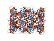
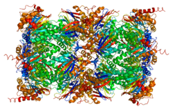
The proteasome is a multicatalytic proteinase complex with a highly ordered 20S core structure. This barrel-shaped core structure is composed of 4 axially stacked rings of 28 non-identical subunits: the two end rings are each formed by 7 alpha subunits, and the two central rings are each formed by 7 beta subunits. Three beta subunits (beta1, beta2, and beta5) each contains a proteolytic active site and has distinct substrate preferences. Proteasomes are distributed throughout eukaryotic cells at a high concentration and cleave peptides in an ATP/ubiquitin-dependent process in a non-lysosomal pathway.[3][4]
Mechanism
Crystal structures of isolated 20S proteasome complex demonstrate that the two rings of beta subunits form a proteolytic chamber and maintain all their active sites of proteolysis within the chamber.[4] Concomitantly, the rings of alpha subunits form the entrance for substrates entering the proteolytic chamber. In an inactivated 20S proteasome complex, the gate into the internal proteolytic chamber are guarded by the N-terminal tails of specific alpha-subunit.[5][6] The proteolytic capacity of 20S core particle (CP) can be activated when CP associates with one or two regulatory particles (RP) on one or both side of alpha rings. These regulatory particles include 19S proteasome complexes, 11S proteasome complex, etc. Following the CP-RP association, the confirmation of certain alpha subunits will change and consequently cause the opening of substrate entrance gate. Besides RPs, the 20S proteasomes can also be effectively activated by other mild chemical treatments, such as exposure to low levels of sodium dodecylsulfate (SDS) or NP-14.[6] [7]
Interactions
PSMA5 has been shown to interact with PLK1.[8]
References
- ↑ DeMartino GN, Orth K, McCullough ML, Lee LW, Munn TZ, Moomaw CR et al. (Oct 1991). "The primary structures of four subunits of the human, high-molecular-weight proteinase, macropain (proteasome), are distinct but homologous". Biochim Biophys Acta 1079 (1): 29–38. doi:10.1016/0167-4838(91)90020-Z. PMID 1888762.
- ↑ 2.0 2.1 "Entrez Gene: PSMA5 proteasome (prosome, macropain) subunit, alpha type, 5".
- ↑ Coux O, Tanaka K, Goldberg AL (1996). "Structure and functions of the 20S and 26S proteasomes". Annual Review of Biochemistry 65: 801–47. doi:10.1146/annurev.bi.65.070196.004101. PMID 8811196.
- ↑ 4.0 4.1 Tomko RJ, Hochstrasser M (2013). "Molecular architecture and assembly of the eukaryotic proteasome". Annual Review of Biochemistry 82: 415–45. doi:10.1146/annurev-biochem-060410-150257. PMID 23495936.
- ↑ Groll M, Ditzel L, Löwe J, Stock D, Bochtler M, Bartunik HD et al. (Apr 1997). "Structure of 20S proteasome from yeast at 2.4 A resolution". Nature 386 (6624): 463–71. doi:10.1038/386463a0. PMID 9087403.
- ↑ 6.0 6.1 Groll M, Bajorek M, Köhler A, Moroder L, Rubin DM, Huber R et al. (Nov 2000). "A gated channel into the proteasome core particle". Nature Structural Biology 7 (11): 1062–7. doi:10.1038/80992. PMID 11062564.
- ↑ Zong C, Gomes AV, Drews O, Li X, Young GW, Berhane B et al. (Aug 2006). "Regulation of murine cardiac 20S proteasomes: role of associating partners". Circulation Research 99 (4): 372–80. doi:10.1161/01.RES.0000237389.40000.02. PMID 16857963.
- ↑ Feng Y, Longo DL, Ferris DK (Jan 2001). "Polo-like kinase interacts with proteasomes and regulates their activity". Cell Growth Differ. 12 (1): 29–37. PMID 11205743.
Further reading
- Coux O, Tanaka K, Goldberg AL (1996). "Structure and functions of the 20S and 26S proteasomes.". Annu. Rev. Biochem. 65: 801–47. doi:10.1146/annurev.bi.65.070196.004101. PMID 8811196.
- Goff SP (2003). "Death by deamination: a novel host restriction system for HIV-1.". Cell 114 (3): 281–3. doi:10.1016/S0092-8674(03)00602-0. PMID 12914693.
- Rasmussen HH, van Damme J, Puype M, Gesser B, Celis JE, Vandekerckhove J (1993). "Microsequences of 145 proteins recorded in the two-dimensional gel protein database of normal human epidermal keratinocytes.". Electrophoresis 13 (12): 960–9. doi:10.1002/elps.11501301199. PMID 1286667.
- Kristensen P, Johnsen AH, Uerkvitz W, Tanaka K, Hendil KB (1995). "Human proteasome subunits from 2-dimensional gels identified by partial sequencing.". Biochem. Biophys. Res. Commun. 205 (3): 1785–9. doi:10.1006/bbrc.1994.2876. PMID 7811265.
- Maruyama K, Sugano S (1994). "Oligo-capping: a simple method to replace the cap structure of eukaryotic mRNAs with oligoribonucleotides.". Gene 138 (1–2): 171–4. doi:10.1016/0378-1119(94)90802-8. PMID 8125298.
- Seeger M, Ferrell K, Frank R, Dubiel W (1997). "HIV-1 tat inhibits the 20 S proteasome and its 11 S regulator-mediated activation". J. Biol. Chem. 272 (13): 8145–8. doi:10.1074/jbc.272.13.8145. PMID 9079628.
- Suzuki Y, Yoshitomo-Nakagawa K, Maruyama K, Suyama A, Sugano S (1997). "Construction and characterization of a full length-enriched and a 5'-end-enriched cDNA library". Gene 200 (1–2): 149–56. doi:10.1016/S0378-1119(97)00411-3. PMID 9373149.
- Mayau V, Baron B, Buttin G, Debatisse M (1998). "Twelve genes, including the unassigned proteasome zeta subunit gene, ordered within the human 1p13 region". Mamm. Genome 9 (4): 331–3. doi:10.1007/s003359900761. PMID 9530635.
- Madani N, Kabat D (1998). "An endogenous inhibitor of human immunodeficiency virus in human lymphocytes is overcome by the viral Vif protein". J. Virol. 72 (12): 10251–5. PMC 110608. PMID 9811770.
- Simon JH, Gaddis NC, Fouchier RA, Malim MH (1998). "Evidence for a newly discovered cellular anti-HIV-1 phenotype". Nat. Med. 4 (12): 1397–400. doi:10.1038/3987. PMID 9846577.
- Jørgensen L, Hendil KB (1999). "Proteasome subunit zeta, a putative ribonuclease, is also found as a free monomer". Mol. Biol. Rep. 26 (1–2): 119–23. doi:10.1023/A:1006965602142. PMID 10363657.
- Mulder LC, Muesing MA (2000). "Degradation of HIV-1 integrase by the N-end rule pathway". J. Biol. Chem. 275 (38): 29749–53. doi:10.1074/jbc.M004670200. PMID 10893419.
- Feng Y, Longo DL, Ferris DK (2001). "Polo-like kinase interacts with proteasomes and regulates their activity". Cell Growth Differ. 12 (1): 29–37. PMID 11205743.
- Engidawork E, Juranville JF, Fountoulakis M, Dierssen M, Lubec G (2002). "Selective upregulation of the ubiquitin-proteasome proteolytic pathway proteins, proteasome zeta chain and isopeptidase T in fetal Down syndrome". J. Neural Transm. Suppl. (61): 117–30. doi:10.1007/978-3-7091-6262-0_10. PMID 11771738.
- Sheehy AM, Gaddis NC, Choi JD, Malim MH (2002). "Isolation of a human gene that inhibits HIV-1 infection and is suppressed by the viral Vif protein". Nature 418 (6898): 646–50. doi:10.1038/nature00939. PMID 12167863.
- Huang X, Seifert U, Salzmann U, Henklein P, Preissner R, Henke W et al. (2002). "The RTP site shared by the HIV-1 Tat protein and the 11S regulator subunit alpha is crucial for their effects on proteasome function including antigen processing". J. Mol. Biol. 323 (4): 771–82. doi:10.1016/S0022-2836(02)00998-1. PMID 12419264.
- Gaddis NC, Chertova E, Sheehy AM, Henderson LE, Malim MH (2003). "Comprehensive investigation of the molecular defect in vif-deficient human immunodeficiency virus type 1 virions". J. Virol. 77 (10): 5810–20. doi:10.1128/JVI.77.10.5810-5820.2003. PMC 154025. PMID 12719574.
| |||||||||