Optic vesicle
| Optic vesicle | |
|---|---|
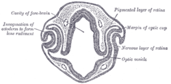
 Transverse section of head of chick embryo of forty-eight hours’ incubation. (Optic vesicle labeled at lower right.) | |
 | |
| Details | |
| Latin | vesicula optica; vesicula ophthalmica |
| Carnegie stage | 11 |
| Identifiers | |
| Gray's | p.1001 |
| Code | TE E5.14.3.4.2.2.4 |
| Anatomical terminology | |
The eyes begin to develop as a pair of diverticula from the lateral aspects of the forebrain. These diverticula make their appearance before the closure of the anterior end of the neural tube; after the closure of the tube they are known as the optic vesicles.
They project toward the sides of the head, and the peripheral part of each expands to form a hollow bulb, while the proximal part remains narrow and constitutes the optic stalk.
Additional images
-

Head of chick embryo of about thirty-eight hours’ incubation, viewed from the ventral surface. X 26
References
This article incorporates text in the public domain from the 20th edition of Gray's Anatomy (1918)
External links
- eye-012 — Embryo Images at University of North Carolina
- Overview at vision.ca
- Overview at temple.edu
| ||||||||||||||||||||||||||||||||||||||||||||||||||||||||||||||||||||||