Occipital horn of lateral ventricle
| Occipital horn of lateral ventricle | |
|---|---|
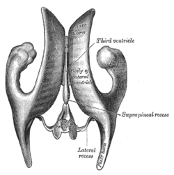
 Drawing of a cast of the ventricular cavities, viewed from above. | |
 Drawing of a cast of the ventricular cavities, viewed from the side. | |
| Details | |
| Latin | cornu posterius |
| Identifiers | |
| Gray's | p.829 |
| NeuroNames | hier-205 |
| NeuroLex ID | Posterior horn lateral ventricle |
| TA | A14.1.09.286 |
| FMA | 83700 |
| Anatomical terms of neuroanatomy | |
The occipital horn of the lateral ventricle posteriorly (also posterior cornu of the lateral ventricle, postcornu of the lateral ventricle) passes into the occipital lobe, its direction being backward and lateralward, and then medialward.
Its roof is formed by the fibers of the corpus callosum passing to the temporal and occipital lobes.
On its medial wall is a longitudinal eminence, the calcar avis (hippocampus minor), which is an involution of the ventricular wall produced by the calcarine fissure.
Above this the forceps posterior of the corpus callosum, sweeping around to enter the occipital lobe, causes another projection, termed the bulb of the posterior cornu.
The calcar avis and bulb of the posterior cornu are extremely variable in their degree of development; in some cases they are ill-defined, in others prominent.
Additional images
-
Human brain right dissected lateral view
-
Ventricles of braian and basal ganglia.Superior view. Horizontal section.Deep dissection
-
Ventricles of braian and basal ganglia.Superior view. Horizontal section.Deep dissection
References
This article incorporates text in the public domain from the 20th edition of Gray's Anatomy (1918)
External links
- Atlas image: n1a4p2 at the University of Michigan Health System
- Atlas image: n1a4p5 at the University of Michigan Health System
| ||||||||||||||||||||||||||||


