Myoglobin
| Myoglobin | |||||||||||||
|---|---|---|---|---|---|---|---|---|---|---|---|---|---|
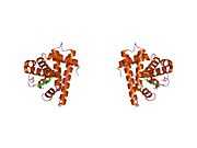
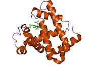
 Model of helical domains in myoglobin.[1] | |||||||||||||
| |||||||||||||
| Identifiers | |||||||||||||
| Symbols | MB ; PVALB | ||||||||||||
| External IDs | OMIM: 160000 MGI: 96922 HomoloGene: 3916 GeneCards: MB Gene | ||||||||||||
| |||||||||||||
| RNA expression pattern | |||||||||||||
 | |||||||||||||
| More reference expression data | |||||||||||||
| Orthologs | |||||||||||||
| Species | Human | Mouse | |||||||||||
| Entrez | 4151 | 17189 | |||||||||||
| Ensembl | ENSG00000198125 | ENSMUSG00000018893 | |||||||||||
| UniProt | P02144 | P04247 | |||||||||||
| RefSeq (mRNA) | NM_005368 | NM_001164047 | |||||||||||
| RefSeq (protein) | NP_005359 | NP_001157519 | |||||||||||
| Location (UCSC) | Chr 22: 36 – 36.03 Mb | Chr 15: 77.02 – 77.05 Mb | |||||||||||
| PubMed search | |||||||||||||
Myoglobin is an iron- and oxygen-binding protein found in the muscle tissue of vertebrates in general and in almost all mammals. It is related to hemoglobin, which is the iron- and oxygen-binding protein in blood, specifically in the red blood cells. In humans, myoglobin is only found in the bloodstream after muscle injury. It is an abnormal finding, and can be diagnostically relevant when found in blood. [2]
Myoglobin is the primary oxygen-carrying pigment of muscle tissues.[3] High concentrations of myoglobin in muscle cells allow organisms to hold their breath for a longer period of time. Diving mammals such as whales and seals have muscles with particularly high abundance of myoglobin.[2] Myoglobin is found in Type I muscle, Type II A and Type II B, but most texts consider myoglobin not to be found in smooth muscle.
Myoglobin was the first protein to have its three-dimensional structure revealed by X-ray crystallography.[4] This achievement was reported in 1958 by John Kendrew and associates.[5] For this discovery, John Kendrew shared the 1962 Nobel Prize in chemistry with Max Perutz.[6] Despite being one of the most studied proteins in biology, its physiological function is not yet conclusively established: mice genetically engineered to lack myoglobin are viable, but showed a 30% reduction in volume of blood being pumped by the heart during a contraction. They adapted to this deficiency through natural reactions to inadequate oxygen supply (hypoxia) and a widening of blood vessels (vasodilation).[7] In humans myoglobin is encoded by the MB gene.[8]
Meat color
Myoglobin contains hemes, pigments responsible for the color of red meat. The color that meat takes is partly determined by the degree of oxidation of the myoglobin. In fresh meat the iron atom is the ferrous state bound to a dioxygen molecule (O2). Meat cooked well done is brown because the iron atom is now in the ferric (+3) oxidation state, having lost an electron. If meat has been exposed to nitrites, it will remain pink because the iron atom is bound to NO, nitric oxide (true of, e.g., corned beef or cured hams). Grilled meats can also take on a pink "smoke ring" that comes from the iron binding to a molecule of carbon monoxide.[9] Raw meat packed in a carbon monoxide atmosphere also shows this same pink "smoke ring" due to the same principles. Notably, the surface of this raw meat also displays the pink color, which is usually associated in consumers' minds with fresh meat. This artificially induced pink color can persist, reportedly up to one year.[10] Hormel and Cargill are both reported to use this meat-packing process, and meat treated this way has been in the consumer market since 2003.[11]
Role in disease
Myoglobin is released from damaged muscle tissue (rhabdomyolysis), which has very high concentrations of myoglobin. The released myoglobin is filtered by the kidneys but is toxic to the renal tubular epithelium and so may cause acute renal failure.[12] It is not the myoglobin itself that is toxic (it is a protoxin) but the ferrihemate portion that is dissociated from myoglobin in acidic environments (e.g., acidic urine, lysosomes).
Myoglobin is a sensitive marker for muscle injury, making it a potential marker for heart attack in patients with chest pain.[13] However, elevated myoglobin has low specificity for acute myocardial infarction (AMI) and thus CK-MB, cTnT, ECG, and clinical signs should be taken into account to make the diagnosis.
Structure and bonding

Myoglobin belongs to the globin superfamily of proteins, and as with other globins, consists of eight alpha helices connected by loops. Human globin contains 154 amino acids.[15]
Myoglobin contains a porphyrin ring with an iron at its center. A proximal histidine group (His-94) is attached directly to iron, and a distal histidine group (His-65) hovers near the opposite face.[15] The distal imidazole is not bonded to the iron but is available to interact with the substrate O2. This interaction encourages the binding of O2, but not carbon monoxide (CO), which still binds about 240× more strongly than O2.
The binding of O2 causes substantial structural change at the Fe center, which shrinks in radius and moves into the center of N4 pocket. O2-binding induces "spin-pairing": the five-coordinate ferrous deoxy form is high spin and the six coordinate oxy form is low spin and diamagnetic.
Synthetic analogues
Many models of myoglobin have been synthesized as part of a broad interest in transition metal dioxygen complexes. A well known example is the picket fence porphyrin, which consists of a ferrous complex of a sterically bulky derivative of tetraphenylporphyrin.[16] In the presence of an imidazole ligand, this ferrous complex reversibly binds O2. The O2 substrate adopts a bent geometry, occupying the sixth position of the iron center. A key property of this model is the slow formation of the μ-oxo dimer, which is an inactive diferric state. In nature, such deactivation pathways are suppressed by protein matrix that prevents close approach of the Fe-porphyrin assemblies.[17]
 A picket-fence porphyrin complex of Fe, with axial coordination sites occupied by methylimidazole (green) and dioxygen. The R groups flank the O2-binding site.
See also
- Cytoglobin
- Hemoglobin
- Hemoprotein
- Neuroglobin
References
- ↑ PDB 1MBO; Takano T (March 1977). "Structure of myoglobin refined at 2.0 Å resolution. II. Structure of deoxymyoglobin from sperm whale". J. Mol. Biol. 110 (3): 569–84. doi:10.1016/S0022-2836(77)80112-5. PMID 845960.
- ↑ 2.0 2.1 Nelson DL, Cox MM (2000). Lehninger Principles of Biochemistry (3rd ed.). New York: Worth Publishers. p. 206. ISBN 0-7167-6203-X. (Google books link is the 2008 edition)
- ↑ Ordway GA, Garry DJ (September 2004). "Myoglobin: an essential hemoprotein in striated muscle". J. Exp. Biol. 207 (Pt 20): 3441–6. doi:10.1242/jeb.01172. PMID 15339940.
- ↑ (U.S.) National Science Foundation: Protein Data Bank Chronology (Jan. 21, 2004). Retrieved 3.17.2010
- ↑ Kendrew JC, Bodo G, Dintzis HM, Parrish RG, Wyckoff H, Phillips DC (1958). "A Three-Dimensional Model of the Myoglobin Molecule Obtained by X-Ray Analysis". Nature 181 (4610): 662–6. Bibcode:1958Natur.181..662K. doi:10.1038/181662a0. PMID 13517261.
- ↑ The Nobel Prize in Chemistry 1962
- ↑ Mammen PP, Kanatous SB, Yuhanna IS, Shaul PW, Garry MG, Balaban RS, Garry DJ (2003). "Hypoxia-induced left ventricular dysfunction in myoglobin-deficient mice". American Journal of Physiology. Heart and Circulatory Physiology 285 (5): H2132–41. doi:10.1152/ajpheart.00147.2003. PMID 12881221.
- ↑ Akaboshi E (1985). "Cloning of the human myoglobin gene". Gene 33 (3): 241–9. doi:10.1016/0378-1119(85)90231-8. PMID 2989088.
- ↑ McGee H (2004). On Food and Cooking: The Science and Lore of the Kitchen. New York: Scribner. p. 148. ISBN 0-684-80001-2.
- ↑ Fraqueza MJ, Barreto AS (September 2011). "Gas mixtures approach to improve turkey meat shelf life under modified atmosphere packaging: the effect of carbon monoxide". Poult. Sci. 90 (9): 2076–84. doi:10.3382/ps.2011-01366. PMID 21844276.
- ↑ Associated Press (2007-10-30). "Meat companies defend use of carbon monoxide". Business. Minneapolis Star Tribune.
- ↑ Naka T, Jones D, Baldwin I, Fealy N, Bates S, Goehl H, Morgera S, Neumayer HH, Bellomo R (April 2005). "Myoglobin clearance by super high-flux hemofiltration in a case of severe rhabdomyolysis: a case report". Crit Care 9 (2): R90–5. doi:10.1186/cc3034. PMC 1175920. PMID 15774055.
- ↑ Weber M, Rau M, Madlener K, Elsaesser A, Bankovic D, Mitrovic V, Hamm C (November 2005). "Diagnostic utility of new immunoassays for the cardiac markers cTnI, myoglobin and CK-MB mass". Clin. Biochem. 38 (11): 1027–30. doi:10.1016/j.clinbiochem.2005.07.011. PMID 16125162.
- ↑ Drago RS (1980). "Free radical reactions of transition metal systems". Coordination Chemistry Reviews 32 (2): 97–110. doi:10.1016/S0010-8545(00)80372-0.
- ↑ 15.0 15.1 UniProt: P02144
- ↑ Collman JP, Brauman JI, Halbert TR, Suslick KS (October 1976). "Nature of O2 and CO binding to metalloporphyrins and heme proteins". Proc. Natl. Acad. Sci. U.S.A. 73 (10): 3333–7. Bibcode:1976PNAS...73.3333C. doi:10.1073/pnas.73.10.3333. PMC 431107. PMID 1068445.
- ↑ Lippard SJ, Berg JM (1994). Principles of Bioinorganic Chemistry. Mill Valley, CA: University Science Books. ISBN 0-935702-73-3.
Further reading
- Collman JP, Boulatov R, Sunderland CJ, Fu L (February 2004). "Functional analogues of cytochrome c oxidase, myoglobin, and hemoglobin". Chem. Rev. 104 (2): 561–88. doi:10.1021/cr0206059. PMID 14871135.
- Reeder BJ, Svistunenko DA, Cooper CE, Wilson MT (December 2004). "The radical and redox chemistry of myoglobin and hemoglobin: from in vitro studies to human pathology". Antioxid. Redox Signal. 6 (6): 954–66. doi:10.1089/ars.2004.6.954. PMID 15548893.
- Schlieper G, Kim JH, Molojavyi A, Jacoby C, Laussmann T, Flögel U, Gödecke A, Schrader J (April 2004). "Adaptation of the myoglobin knockout mouse to hypoxic stress". Am. J. Physiol. Regul. Integr. Comp. Physiol. 286 (4): R786–92. doi:10.1152/ajpregu.00043.2003. PMID 14656764.
- Takano T (1977). "Structure of myoglobin refined at 2.0 Å resolution. II. Structure of deoxymyoglobin from sperm whale". J. Mol. Biol. 110 (3): 569–584. doi:10.1016/S0022-2836(77)80112-5. PMID 845960.
- Roy A, Sen S, Chakraborti AS (February 2004). "In vitro nonenzymatic glycation enhances the role of myoglobin as a source of oxidative stress". Free Radic. Res. 38 (2): 139–46. doi:10.1080/10715160310001638038. PMID 15104207.
- Stewart JM, Blakely JA, Karpowicz PA, Kalanxhi E, Thatcher BJ, Martin BM (March 2004). "Unusually weak oxygen binding, physical properties, partial sequence, autoxidation rate and a potential phosphorylation site of beluga whale (Delphinapterus leucas) myoglobin". Comp. Biochem. Physiol. B, Biochem. Mol. Biol. 137 (3): 401–12. doi:10.1016/j.cbpc.2004.01.007. PMID 15050527.
- Wu G, Wainwright LM, Poole RK (2003). "Microbial globins". Adv. Microb. Physiol. Advances in Microbial Physiology 47: 255–310. doi:10.1016/S0065-2911(03)47005-7. ISBN 9780120277476. PMID 14560666.
External links
- The Myoglobin Protein
- Protein Database featured molecule
- Online 'Mendelian Inheritance in Man' (OMIM) 160000 human genetics
- Which Cut Is Older? (It's a Trick Question) New York Times, February 21, 2006 article regarding meat industry use of carbon monoxide to keep meat looking red.
- Stores React to Meat Reports New York Times, March 1, 2006 article on the use of carbon monoxide to make meat appear fresh.
- Mirceta, S.; Signore, A. V.; Burns, J. M.; Cossins, A. R.; Campbell, K. L.; Berenbrink, M. (2013). "Evolution of Mammalian Diving Capacity Traced by Myoglobin Net Surface Charge". Science 340 (6138): 1234192. doi:10.1126/science.1234192. PMID 23766330.. Also see Proteopedia article about this finding
| |||||||||||||||||||||||||||||||||||||||||||


| ||||||||||||||||||||||||||||||||||||||||||||||||||||||||||||||||||||||||||||||||
| ||||||||||||||||||||||||||||||||||||||||||||||||||||||||||||||||||||||||||||||||||||||||||||||