mir-19 microRNA precursor family
| mir-19 microRNA precursor family | |
|---|---|
 | |
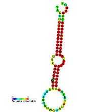
| Predicted secondary structure and sequence conservation of mir-19 | |
| Identifiers | |
| Symbol | mir-19 |
| Rfam | RF00245 |
| miRBase | MI0000073 |
| miRBase family | MIPF0000011 |
| Other data | |
| RNA type | Gene; miRNA |
| Domain(s) | Eukaryota |
| GO | 0035195 0035068 |
| SO | 0001244 |
There are 89 known sequences today in the microRNA 19 (miR-19) family but it will change quickly. They are found in a large number of vertebrate species. The miR-19 microRNA precursor is a small non-coding RNA molecule that regulates gene expression. Within the human and mouse genome there are three copies of this microRNA that are processed from multiple predicted precursor hairpins:[1][2][3]
- mouse:
- * miR-19a on chromosome 14 (MI0000688)
- * miR-19b-1 on chromosome 14 (MI0000718)
- * miR-19b-2 on chromosome X (MI0000546)
- human:[1]
- * miR-19a on chromosome 13 (MI0000073)
- * miR-19b-1 on chromosome 13 (MI0000074)
- * miR-19b-2 on chromosome X (MI000075).
MiR-19 has now been predicted or experimentally confirmed (MIPF0000011). In this case the mature sequence is excised from the 3' arm of the hairpin precursor.
Origins
MicroRNA are ubiquitous in higher eukaryotes, and show varying patterns of expression in specific cell types.[4] MiR-19 has been identified in a diverse range of vertebrate animals including green anole (Anolis carolinensis),[5] primates (gorilla, human,…),[6][7] cattle (Bos taurus),[8] dog,[9] chinese hamster (Cricetulus griseus),[10] zebrafish (Danio rerio),[11] horse (Equus caballus),[12] Takifugu rubripes,[11]Tetraodon nigroviridis,[11] chicken (Gallus gallus),[13][14] gray short-tailed opossum (Monodelphis domestica),[15] platypus (Ornithorhynchus anatinus),[16] Japanese medaka (Oryzias latipes),[17] Xenopus laevis (frog),[18] Tasmanian devil (Sarcophilus harrisii),[19] pig (Sus scrofa)[20] and Zebra Finch (Taeniopygia guttata).[21] In some of these species the presence of miR-19 microRNAs have been directly measured, in other species genes have been identified with sequences that are predicted to encode miR-19.[1]
Expression
MiR-17-92 cluster was identified to encode 6 single mature miRNA (miR-17, , miR-19, miR-20, miR-92, miR-106) containing the first oncogenic miRNA.
MicroRNA from miR-19 family can be expressed from:
- * T-cell acute lymphoblastic leukemia [22]
- * B-cell lymphomas [23]
- * Cell lines [22]
- * Cerebellum [24][25]
- * Purkinje cells [24]
- * HeLa cells [26]
Finally they have tissues-specific miRNA expression. These microRNA are considered as oncogenes which improve proliferation, inhibits apoptosis and induce tumor angiogenesis.[27]
These miRNA are context-specific and they have different roles depending on where they are.
miR-19a/b roles
Acute lymphoblastic leukemia
Ectopic expression of miR-19 represses CYLD expression, while miR-19 inhibitor treatment induces CYLD protein expression and decreases NF-kB expression in the downstream signaling pathway.
Thus, miR-19, CYLD and NF-kB form a regulatory feedforward loop, which provides new clues for sustained activation of NF-kB in T-cell acute lymphoblastic leukemia.[22]
MiR-19 is sufficient to induce T-cell lymphoblastic leukemia activating Notch1 and accelerate the disease. Its targets are:
- * Bim (Bcl2L11) gene
- * AMP-activated kinase (Prkaa1) gene
- * E2F1 gene
- * the tumour suppressor phosphatases PTEN
- * PP2A (Ppp2r5e) gene
- * Dock5 protein
MiR-19b coordinates a PI3K pathway acting on cell survival in lymphocytes contributing to leukaemogenesis.[28][29][30]
This pathway is activated through PTEN loss and can contribute to reduce sensitivity to chemotherapy and (in T-ALL) may impact the effectiveness of therapeutic gamma-secretase inhibitors.
Primary central nervous system lymphoma
Baraniskin and al. study show that miR-21, miR-19, and miR-92a levels in cerebrospinal fluid (CSF) seems to be good biomarkers to diagnose a Primary central nervous system lymphoma (PCNSL). They also demonstrate that miRNAs in plasma are in a resistant form to intrinsic RNase activity, and there is a low RNase activity in the CSF.[25]
B-cell lymphomas
MiR-19 has been identified as a key responsible for the oncogenic activity, reducing the tumor suppressor gene PTEN expression and activating AKT/mTOR pathway. This cluster might be important regulator on cancer and aging.[31][32]
Mu and al. demonstrated that the expression of endogenous miR-17-92 is required to suppress apoptosis in Myc-driven B-cell lymphomas. More specifically, miR-19a and miR-19b are required and sufficient to recapitulate the oncogenic properties of the entire cluster.[23][33]
Using prediction algorithms, they found miR-19 targets to the pro-survival functions:
Keratinocytes
In the cell response to stress, the most important is the post-transcriptional control of the important gene expression to cell survival and apoptosis. MiR-19 regulates the Ras homolog B (RhoB) expression in keratinocytes after ultraviolet (UV) radiation exposition. This phenomenon needs the binding of human antigen R (HuR) to the rhoB mRNA 3'-untranslated region. In this case, HuR acts positively on miRNA action. The interaction between HuR and miR-19 with rhoB is lost under UV treatment. Here, miR-19, linked to RhoB, acts like a protector against keratinocyte apoptosis. A 52-nucleotide-long sequence of the rhoB 3'-UTR spanning bases 818–870, containing the miR-19 and the HuR binding site was sufficient for UV regulation. This event is UV dependent![34]
Multiple Myeloma (MM)
One study on multiple myeloma patients permitted to identified a selective up-regulation of miR-32 and the miR-17-92 cluster. MiR-19a and miR-19b were shown to down regulate SOCS-1 expression (a specific gene that inhibits IL-6 growth signaling). Therefore, miR-17-92 with miR-21, inhibits apoptosis and promotes cell survival.[33]
Retinoblastoma
In this case, miR-17-92 cluster promotes retinoblastoma due to loss of Rb family members. The mouse retinal development need miR-17-92 over-expresson with Rb and p107 deletion, but it occurred frequent emergence of retinoblastoma and metastasis to the brain.
Here, the cluster oncogenic function is not mediated by a miR-19/PTEN axis toward apoptosis suppression like in lymphoma or in leukemia models. MiR-17-92 increase the proliferative capacity of Rb/p107-deficient in retinal cells.
Moreover, the Rb family members deletion led to compensatory up-regulation of the cyclin-dependent kinase inhibitor p21Cip1.
Finally, the cluster over-expression counteracted p21Cip1 up-regulation, promotes proliferation and drove retinoblastoma formation.[35]
Role in normal development of heart, lungs and immune system
Scientists observed that the loss of function of the miR-17-92 cluster is induced in smaller embryos and postnatal deaths.[36] The specific role of this cluster in heart and lung development remains unclear, but the observations described above show that these miRNAs are normally highly expressed in embryonic lung and decrease with maturity. Moreover, transgenic expression of these miRNAs specifically in lung epithelium results in severe developmental defects with enhanced proliferation and
inhibition of differentiation of epithelial cells.
Furthermore, mouse hematopoiesis occurring in the absence of miR-17-92 leads to an isolated defect in B cell development.[36]
Role in the endothelial differentiation of stem cells
The miR-17-92 cluster containing miR-19 miRNA family is also involved into control endothelial cell functions and neo-vascularization. MiRNA cluster (miR-17, miR-18, miR-19 and miR-20) increased during the induction of endothelial cell differentiation in embryonic stem cells (tested on murine) or induce pluripotent stem cells. Even though this cluster regulates vascular integrity and angiogenesis, none of each members has a significant impact on the endothelial differentiation of pluripotent stem cells.[37]
miR-19a Roles
Spinocerebellar ataxia type 1
It has been showing that the 3' UTR of the ATXN1 gene contains 3 target sites for miR-19, and this microRNA shows moderate down regulation of reporter genes containing the ATXN1 3' UTR. Furthermore, it directly binds to the ATXN1 3´UTR to suppress the translation of ATXN1. ATXN1 is also regulated by miR-101, and miR-130.[24]
Breast Cancer
MiR-19 regulates tissue factor expression at a post-transcriptional level in breast cancer cells, providing a molecular basis for the selective expression of the tissue factor gene. Thanks to bioinformatics analyses, scientists predicted microRNA-Binding sites for miR-19, miR-20 and miR-106b in the 3'-UTR tissue factor transcript. Experiments confirmed that it negatively regulates gene expression in MCF-7 cells, and over-expression of miR-19 downregulates tissue factor expression in MDA-MB-231 cells (Human breast cancer cell lines). The main action of miR-19 seems to inhibit protein translation of the tissue factor gene in less invasive breast cancer cells.[27]
miR-19b Roles
Rheumatoid arthritis
MiR-19 also takes part in inflammatory responses enhancing or repressing pro-inflammatory mediators expression. It positively regulates Toll-like receptor signaling with Dicer1 deletion and miRNA depletion. MiR-19b is an important protagonist in this phenomenon, regulating positively NF-kB activity.
MiRNA depletion inhibits cytokines production by NF-kB. This indicates that miRNA control of NF-kB signaling repressors thanks to its relief. Some important regulators of NF-kB signaling (like A20 (Tnfaip3), Cyld, and Cezanne (Otud7b)) is targeted by the miR-17-92 cluster.
Moreover, mir-19 targets some members of the Tnfaip3-ubiquitin editing complex (Tnfaip3/Itch/Tnip1/Rnf11). MiR-19 directly involved in the modulation of several NF-kB signaling negative regulators expression, indicating an important role for Rnf11 in the effect of miR-19b on NF-kB signaling.
Finally, miR-19b exacerbates the cells crucial inflammatory activation in rheumatoid arthritis disease.[26][29]
References
- ↑ 1.0 1.1 1.2 Lagos-Quintana, M; Rauhut R; Lendeckel W; Tuschl T (2001). "Identification of novel genes coding for small expressed RNAs". Science 294 (5543): 853–858. doi:10.1126/science.1064921. PMID 11679670.
- ↑ Mourelatos, Z; Dostie J; Paushkin S; Sharma A; Charroux B; Abel L; Rappsilber J; Mann M; Dreyfuss G (2002). "miRNPs: a novel class of ribonucleoproteins containing numerous microRNAs". Genes Dev 16 (6): 720–728. doi:10.1101/gad.974702. PMC 155365. PMID 11914277.
- ↑ Houbaviy, HB; Murray MF; Sharp PA (2003). "Embryonic stem cell-specific MicroRNAs". Dev Cell 5 (2): 351–358. doi:10.1016/S1534-5807(03)00227-2. PMID 12919684.
- ↑ Landgraf, P; M Rusu; R Sheridan; A Sewer (2007). "A Mammalian microRNA Expression Atlas Based on Small RNA Library Sequencing". Cell. 129 (7): 1401–1414. doi:10.1016/j.cell.2007.04.040. PMC 2681231. PMID 17604727.
- ↑ Lyson TR, Sperling EA, Heimberg AM and al. (2012). "MicroRNAs support a turtle + lizard clade". Biol Lett 8 (1): 104–7. doi:10.1098/rsbl.2011.0477. PMC 3259949. PMID 21775315.
- ↑ Berezikov E, Guryev V, van de Belt J and al. (2005). "Phylogenetic shadowing and computational identification of human microRNA genes". Cell 120 (1): 21–4. doi:10.1016/j.cell.2004.12.031. PMID 15652478.
- ↑ Lui WO, Pourmand N, Patterson BK and al. (2007). "Patterns of known and novel small RNAs in human cervical cancer". Cancer Res 67 (13): 6031–43. doi:10.1158/0008-5472.CAN-06-0561. PMID 17616659.
- ↑ Gu Z, Eleswarapu S, Jiang H (2007). "Identification and characterization of microRNAs from the bovine adipose tissue and mammary gland". FEBS Lett 581 (5): 981–8. doi:10.1016/j.febslet.2007.01.081. PMID 17306260.
- ↑ Friedländer MR, Chen W, Adamidi C and al. (2008). "Discovering microRNAs from deep sequencing data using miRDeep". Nat Biotechnol 26 (4): 407–15. doi:10.1038/nbt1394. PMID 18392026.
- ↑ Hackl M, Jakobi T, Blom J and al. (2011). "Next-generation sequencing of the Chinese hamster ovary microRNA transcriptome: Identification, annotation and profiling of microRNAs as targets for cellular engineering". J Biotechnol 153 (1-2): 62–75. doi:10.1016/j.jbiotec.2011.02.011. PMC 3119918. PMID 21392545.
- ↑ 11.0 11.1 11.2 Chen PY, Manninga H, Slanchev K and al. (2005). "The developmental miRNA profiles of zebrafish as determined by small RNA cloning". Genes Dev 19 (11): 1288–93. doi:10.1101/gad.1310605. PMC 1142552. PMID 15937218.
- ↑ Zhou M, Wang Q, Sun J and al. (2009). "In silico detection and characteristics of novel microRNA genes in the Equus caballus genome using an integrated ab initio and comparative genomic approach". Genomics 94 (2): 125–31. doi:10.1016/j.ygeno.2009.04.006. PMID 19406225.
- ↑ International Chicken Genome Sequencing Consortium (2004). "Sequence and comparative analysis of the chicken genome provide unique perspectives on vertebrate evolution". Nature 432 (7018): 695–716. doi:10.1038/nature03154. PMID 15592404.
- ↑ Yao Y, Zhao Y, Xu H and al. (2008). "MicroRNA profile of Marek's disease virus-transformed T-cell line MSB-1: predominance of virus-encoded microRNAs". J Virol 82 (8): 4007–15. doi:10.1128/JVI.02659-07. PMC 2293013. PMID 18256158.
- ↑ Devor EJ, Samollow PB (2008). "In vitro and in silico annotation of conserved and nonconserved microRNAs in the genome of the marsupial Monodelphis domestica". J Hered 99 (1): 66–72. doi:10.1093/jhered/esm085. PMID 17965199.
- ↑ Murchison EP, Kheradpour P, Sachidanandam R and al. (2008). "Conservation of small RNA pathways in platypus". Genome Res 18 (6): 995–1004. doi:10.1101/gr.073056.107. PMC 2413167. PMID 18463306.
- ↑ Li SC, Chan WC, Ho MR and al. (2010). "Discovery and characterization of medaka miRNA genes by next generation sequencing platform". BMC Genomics. 11 Suppl 4: S8. doi:10.1186/1471-2164-11-S4-S8. PMC 3005926. PMID 21143817.
- ↑ Watanabe T, Takeda A, Mise K and al. (2005). "Stage-specific expression of microRNAs during Xenopus development". FEBS Lett 579 (2): 318–24. doi:10.1016/j.febslet.2004.11.067. PMID 15642338.
- ↑ Murchison EP, Tovar C, Hsu A and al. (2010). "The Tasmanian devil transcriptome reveals Schwann cell origins of a clonally transmissible cancer". Science 327 (5961): 84–7. doi:10.1126/science.1180616. PMC 2982769. PMID 20044575.
- ↑ Wernersson R, Schierup MH, Jørgensen FG and al. (2005). "Pigs in sequence space: a 0.66X coverage pig genome survey based on shotgun sequencing". BMC Genomics 6: 6:70. doi:10.1186/1471-2164-6-70. PMC 1142312. PMID 15885146.
- ↑ Warren WC, Clayton DF, Ellegren H and al. (2010). "The genome of a songbird". Nature 464 (7289): 757–62. doi:10.1038/nature08819. PMC 3187626. PMID 20360741.
- ↑ 22.0 22.1 22.2 Huashan Ye, Xiaowen Liu, Meng Lv, Yuliang Wu, Shuzhen Kuang, Jing Gong, Ping Yuan, Zhaodong Zhong, Qiubai Li, Haibo Jia, Jun Sun, Zhichao Chen and An-Yuan Guo (2012). "MicroRNA and transcription factor co-regulatory network analysis reveals miR-19 inhibits CYLD in T-cell acute lymphoblastic leukemia". Nucleic Acids Research 40. doi:10.1093/nar/gks175.
- ↑ 23.0 23.1 Ping Mu, Yoon-Chi Han, Doron Betel, Evelyn Yao, Massimo Squatrito, Paul Ogrodowski, Elisa de Stanchina, Aleco D’Andrea, Chris Sander, Andrea Ventura (2009). "Genetic dissection of the miR-17~92 cluster of microRNAs in Myc-induced B-cell lymphomas". Genes Dev 23 (24): 2806–11. doi:10.1101/gad.1872909. PMC 2800095. PMID 20008931.
- ↑ 24.0 24.1 24.2 Lee Y, Samaco RC, Gatchel JR, Thaller C, Orr HT, Zoghbi HY (October 2008). "miR-19, miR-101 and miR-130 co-regulate ATXN1 levels to potentially modulate SCA1 pathogenesis". Nat. Neurosci. 11 (10): 1137–9. doi:10.1038/nn.2183. PMC 2574629. PMID 18758459.
- ↑ 25.0 25.1 Alexander Baraniskin, Jan Kuhnhenn, Uwe Schlegel, Andrew Chan, Martina Deckert, Ralf Gold, Abdelouahid Maghnouj, Hannah Zöllner, Anke Reinacher-Schick, Wolff Schmiegel, Stephan A. Hahn, Roland Schroers (2011). "Identification of microRNAs in the cerebrospinal fluid as marker for primary diffuse large B-cell lymphoma of the central nervous system". Blood 117 (11): 3140–3146. doi:10.1182/blood-2010-09-308684. PMID 21200023.
- ↑ 26.0 26.1 Michael P. Gantier, H. James Stunden, Claire E. McCoy, Mark A. Behlke, Die Wang, Maria Kaparakis-Liaskos, Soroush T. Sarvestani, Yuan H. Yang, Dakang Xu, Sinéad C. Corr, Eric F. Morand, Bryan R. G. Williams (2012). "A miR-19 regulon that controls NF-iB signaling". Nucleic Acids Research 40 (16): 8048–8058. doi:10.1093/nar/gks521. PMC 3439911. PMID 22684508.
- ↑ 27.0 27.1 Xiaoxi Zhang, Haijun Yu, Jessica R. Lou, Jie Zheng, Hua Zhu, Narcis-Ioan Popescu, Florea Lupu, Stuart E. Lind, and Wei-Qun Ding (2011). "MicroRNA-19 (miR-19) Regulates Tissue Factor Expression in Breast Cancer Cells". The journal of Biological chemistry 286: 1429–1435. doi:10.1074/jbc.M110.146530. PMC 3020751. PMID 21059650.
- ↑ Konstantinos J. Mavrakis1, Andrew L. Wolfe, Elisa Oricchio1, Teresa Palomero and al. (2011). "Genome-wide RNAi screen identifies miR-19 targets in Notchinduced acute T-cell leukaemia (T-ALL)". Nat Cell Biol 12 (4): 372–379. doi:10.1038/ncb2037. PMC 2989719. PMID 20190740.
- ↑ 29.0 29.1 Konstantinos J. Mavrakis and Hans-Guido Wendel (2010). "TargetScreen: an unbiased approach to identify functionally important microRNA targets". Cell Cycle 9 (11): 2080–4. doi:10.4161/cc.9.11.11807. PMID 20505335.
- ↑ Séverine Landais, Sébastien Landry, Philippe Legault and al. (2007). "Oncogenic Potential of the miR-106-363 Cluster and Its Implication in Human T-Cell Leukemia". Cancer Res 67 (12): 5699–707. doi:10.1158/0008-5472.CAN-06-4478. PMID 17575136.
- ↑ Johannes Grillari, Matthias Hackl, Regina Grillari-Voglauer (2010). "miR-17–92 cluster: ups and downs in cancer and aging". Biogerontology 11: 501–506. doi:10.1007/s10522-010-9272-9. PMC 2899009. PMID 20437201.
- ↑ Virginie Olive, Margaux J. Bennett, James C. Walker and al. (2009). "miR-19 is a key oncogenic component of mir-17-92". Genes Dev 23 (24): 2839–49. doi:10.1101/gad.1861409. PMC 2800084. PMID 20008935.
- ↑ 33.0 33.1 Flavia Pichiorri, Sung-Suk Suh, Marco Ladetto and al. (2008). "MicroRNAs regulate critical genes associated with multiple myeloma pathogenesis" 105 (35). pp. 12885–90. doi:10.1073/pnas.0806202105. PMC 2529070. PMID 18728182.
- ↑ V Glorian, G Maillot, S Polès and al. (2011). "HuR-dependent loading of miRNA RISC to the mRNA encoding the Ras-related small GTPase RhoB controls its translation during UV-induced apoptosis". Cell Death and Differentiation 18 (11): 1692–1701. doi:10.1038/cdd.2011.35. PMC 3190107. PMID 21527938.
- ↑ Karina Conkrite, Maggie Sundby, Shizuo Mukai and al. (2011). "miR-17~92 cooperates with RB pathway mutations to promote retinoblastoma" 25 (16). pp. 1734–45. doi:10.1101/gad.17027411. PMC 3165937. PMID 21816922.
- ↑ 36.0 36.1 Joshua T. Mendell (2008). "miRiad roles for the miR-17-92 cluster in development and disease" 133 (2). pp. 217–22. doi:10.1016/j.cell.2008.04.001. PMC 2732113. PMID 18423194.
- ↑ Karine Tréguer, Eva-Marie Heinrich, Kisho Ohtani and al. (2012). "Role of the MicroRNA-17–92 Cluster in the Endothelial Differentiation of Stem Cells" 49 (5). pp. 447–460. doi:10.1159/000339429. PMID 22797777.
Further reading
- Andrea Ventura, Amanda G. Young, Monte M. Winslow and al. (2008). "Targeted deletion reveals essential and overlapping functions of the miR-17~92 family of miRNA clustersMechanical stretch up-regulates microRNA-26a and induces human airway smooth muscle hypertrophy by suppressing glycogen synthase kinase-3β". Cell 132 (5): 875–886. doi:10.1016/j.cell.2008.02.019. PMC 2323338. PMID 18329372.
- Lixin Hong, Maoyi Lai, Michelle Chen and al. (2010). "The miR-17-92 Cluster of microRNAs Confers Tumorigenicity by Inhibiting Oncogene-Induced Senescence". Cancer Res 70 (21): 8547–8557. doi:10.1016/j.cell.2008.02.019. PMC 2970743. PMID 20851997.
- JR-Shiuan Yang, Michael D. Phillips, Doron Betel and al. (2011). "Widespread regulatory activity of vertebrate microRNA* species". RNA 17 (2): 312–26. doi:10.1016/j.cell.2008.02.019. PMC 3022280. PMID 21177881.
- Joost Kluiver, Johan H. Gibcus, Chris Hettinga and al. (2012). "Rapid Generation of MicroRNA Sponges for MicroRNA Inhibition". PLoS ONE 7 (1): e29275. doi:10.1371/journal.pone.0029275. PMC 3253070. PMID 22238599.
External links
| ||||||||||||||||||