Lipoxygenase
| Lipoxygenase | |||||||||
|---|---|---|---|---|---|---|---|---|---|
|
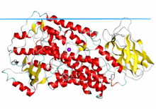
Structure of rabbit reticulocyte 15S-lipoxygenase.[1] | |||||||||
| Identifiers | |||||||||
| Symbol | Lipoxygenase | ||||||||
| Pfam | PF00305 | ||||||||
| InterPro | IPR013819 | ||||||||
| PROSITE | PDOC00077 | ||||||||
| SCOP | 2sbl | ||||||||
| SUPERFAMILY | 2sbl | ||||||||
| OPM superfamily | 87 | ||||||||
| OPM protein | 2p0m | ||||||||
| |||||||||
Lipoxygenases (EC 1.13.11.-) are a family of iron-containing enzymes that catalyze the dioxygenation of polyunsaturated fatty acids in lipids containing a cis,cis-1,4- pentadiene structure. It catalyses the following reaction:
- fatty acid + O2 = fatty acid hydroperoxide
Lipoxygenases are found in plants, animals and fungi. Products of lipoxygenases are involved in diverse cell functions.
Biological function and classification
These enzymes are most common in plants where they may be involved in a number of diverse aspects of plant physiology including growth and development, pest resistance, and senescence or responses to wounding.[2] In mammals a number of lipoxygenases isozymes are involved in the metabolism of eicosanoids (such as prostaglandins, leukotrienes and nonclassic eicosanoids).[3] Sequence data is available for the following lipoxygenases:
- Plant lipoxygenases (EC 1.13.11.12IPR001246). Plants express a variety of cytosolic isozymes as well as what seems to be a chloroplast isozyme.[4]
- Mammalian arachidonate 5-lipoxygenase (EC 1.13.11.34IPR001885).
- Mammalian arachidonate 12-lipoxygenase (EC 1.13.11.31IPR001885).
- Mammalian erythroid cell-specific 15-lipoxygenase (EC 1.13.11.33IPR001885).

3D structure
There are several lipoxygenase structures known including: soybean lipoxygenase L1 and L3, coral 8-lipoxygenase, human 5-lipoxygenase, rabbit 15-lipoxygenase and porcine leukocyte 12-lipoxygenase catalytic domain. The protein consists of a small N-terminal PLAT domain and a major C-terminal catalytic domain (see Pfam link in this article), which contains the active site. In both plant and mammalian enzymes, the N-terminal domain contains an eight-stranded antiparallel β-barrel, but in the soybean lipoxygenases this domain is significantly larger than in the rabbit enzyme. The plant lipoxygenases can be enzymatically cleaved into two fragments which stay tightly associated while the enzyme remains active; separation of the two domains leads to loss of catalytic activity. The C-terminal (catalytic) domain consists of 18-22 helices and one (in rabbit enzyme) or two (in soybean enzymes) antiparallel β-sheets at the opposite end from the N-terminal β-barrel.
Active site
The iron atom in lipoxygenases is bound by four ligands, three of which are histidine residues.[5] Six histidines are conserved in all lipoxygenase sequences, five of them are found clustered in a stretch of 40 amino acids. This region contains two of the three zinc-ligands; the other histidines have been shown[6] to be important for the activity of lipoxygenases.
The two long central helices cross at the active site; both helices include internal stretches of π-helix that provide three histidine (His) ligands to the active site iron. Two cavities in the major domain of soybean lipoxygenase-1 (cavities I and II) extend from the surface to the active site. The funnel-shaped cavity I may function as a dioxygen channel; the long narrow cavity II is presumably a substrate pocket. The more compact mammalian enzyme contains only one boot-shaped cavity (cavity II). In soybean lipoxygenase-3 there is a third cavity which runs from the iron site to the interface of the β-barrel and catalytic domains. Cavity III, the iron site and cavity II form a continuous passage throughout the protein molecule.
The active site iron is coordinated by Nε of three conserved His residues and one oxygen of the C-terminal carboxyl group. In addition, in soybean enzymes the side chain oxygen of asparagine is weakly associated with the iron. In rabbit lipoxygenase, this Asn residue is replaced with His which coordinates the iron via Nδ atom. Thus, the coordination number of iron is either five or six, with a hydroxyl or water ligand to a hexacoordinate iron.
Details about the active site feature of lipoxygenase were revealed in the structure of porcine leukocyte 12-lipoxygenase catalytic domain complex <ref name="PUB00005162.[7] In the 3D structure, the substrate analog inhibitor occupied a U-shaped channel open adjacent to the iron site. This channel could accommodate arachidonic acid without much computation, defining the substrate binding details for the lipoxygenase reaction. In addition, a plausible access channel, which intercepts the substrate binding channel and extended to the protein surface could be counted for the oxygen path.
Biochemical classification
| EC 1.13.11.12 | lipoxygenase | (linoleate:oxygen 13-oxidoreductase) | linoleate + O2 = (9Z,11E,13S)-13-hydroperoxyoctadeca-9,11-dienoate |
| EC 1.13.11.31 | arachidonate 12-lipoxygenase | (arachidonate:oxygen 12-oxidoreductase) | arachidonate + O2 = (5Z,8Z,10E,12S,14Z)-12-hydroperoxyicosa-5,8,10,14-tetraenoate |
| EC 1.13.11.33 | arachidonate 15-lipoxygenase | (arachidonate:oxygen 15-oxidoreductase) | arachidonate + O2 = (5Z,8Z,11Z,13E,15S)-15-hydroperoxyicosa-5,8,11,13-tetraenoate |
| EC 1.13.11.34 | arachidonate 5-lipoxygenase | (arachidonate:oxygen 5-oxidoreductase) | arachidonate + O2 = leukotriene A4 + H2 |
| EC 1.13.11.40 | arachidonate 8-lipoxygenase | (arachidonate:oxygen 8-oxidoreductase) | arachidonate + O2 = (5Z,8R,9E,11Z,14Z)-8-hydroperoxyicosa-5,9,11,14-tetraenoate |
Soybean Lipoxygenase 1 exhibits the largest H/D kinetic isotope effect (KIE) on kcat (kH/kD) (81 near room temperature) so far reported for a biological system.
Human proteins from lipoxygenase family include ALOX12, ALOX12B, ALOX12P2, ALOX15, ALOX15B, ALOX5 and ALOXE3.
References
- ↑ Choi J, Chon JK, Kim S, Shin W (February 2008). "Conformational flexibility in mammalian 15S-lipoxygenase: Reinterpretation of the crystallographic data". Proteins 70 (3): 1023–32. doi:10.1002/prot.21590. PMID 17847087.
- ↑ Vick BA, Zimmerman DC (1987). "Oxidative systems for the modification of fatty acids" 9. pp. 53–90. doi:10.1016/b978-0-12-675409-4.50009-5.
- ↑ Needleman P, Turk J, Jakschik BA, Morrison AR, Lefkowith JB (1986). "Arachidonic acid metabolism". Annu. Rev. Biochem. 55: 69–102. doi:10.1146/annurev.bi.55.070186.000441. PMID 3017195.
- ↑ Tanaka K, Ohta H, Peng YL, Shirano Y, Hibino T, Shibata D (1994). "A novel lipoxygenase from rice. Primary structure and specific expression upon incompatible infection with rice blast fungus". J. Biol. Chem. 269 (5): 3755–3761. PMID 7508918.
- ↑ Boyington JC, Gaffney BJ, Amzel LM (1993). "The three-dimensional structure of an arachidonic acid 15-lipoxygenase". Science 260 (5113): 1482–1486. doi:10.1126/science.8502991. PMID 8502991.
- ↑ Steczko J, Donoho GP, Clemens JC, Dixon JE, Axelrod B (1992). "Conserved histidine residues in soybean lipoxygenase: functional consequences of their replacement". Biochemistry 31 (16): 4053–4057. doi:10.1021/bi00131a022. PMID 1567851.
- ↑ Xu, S.; Mueser T.C.; Marnett L.J.; Funk M.O. (2012). "Crystal structure of 12-lipoxygenase catalytic-domain-inhibitor complex identifies a substrate-binding channel for catalysis.". Structure 20 (9): 1490–7. doi:10.1016/j.str.2012.06.003. PMID 22795085.
External links
- LOX-DB - LipOXygenases DataBase
- Lipoxygenases iron-binding region in PROSITE
- PDB 1YGE - structure of lipoxygenase-1 from soybean (Glycine max)
- PDB 1IK3 - structure of soybean lipoxygenase-3 in complex with (9Z,11E,13S)-13-hydroperoxyoctadeca-9,11-dienoic acid
- PDB 1LOX - structure of rabbit 15-lipoxygenase in complex with inhibitor
- PDB 3RDE - structure of the catalytic domain of porcine leukocyte 12-lipoxygenasean with inhibitor
- UMich Orientation of Proteins in Membranes families/superfamily-87 - animal lipoxygenases
- Lipoxygenase at the US National Library of Medicine Medical Subject Headings (MeSH)
| |||||||||||||||||||||
| ||||||||||
This article incorporates text from the public domain Pfam and InterPro IPR001024
