Leukoaraiosis
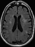
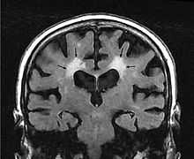
Leukoaraiosis[1] or white matter hyperintensities (WMHs)[2] describes the nonspecific changes in the cerebral white matter frequently seen on CT and MRI in aged individuals and even young adults sometimes.[3] It is a condition routinely found in elderly people.
The term leukoaraiosis was coined in 1986[4][5] as a descriptive term for rarefaction ("araiosis") of the white matter, showing up as decreased density on CT and increased signal intensity on T2/FLAIR sequences (white matter hyperintensities) performed as part of MRI brain scans.
These white matter changes are also commonly referred to as periventricular white matter disease, or white matter hyperintensities (WMH) due to their bright white appearance on T2 MRI scans. Many patients can have leukoaraiosis without any associated clinical abnormality. However, underlying vascular mechanisms are suspected to be the cause of the imaging findings. Hypertension, smoking, diabetes, hyperhomocysteinemia, and heart disease are all risk factors for leukoaraiosis.
Leukoaraiosis has been reported to be an initial stage of Binswanger's disease but this evolution does not always happen.
Causes
White matter hyperintensities can be caused by a variety of factors including ischemia, micro-hemorrhages, gliosis, damage to small blood vessel walls, breaches of the barrier between the cerebrospinal fluid and the brain, or loss and deformation of the myelin sheath.[6] Multiple small vessel infarcts in the subcortical white matter can cause the condition, often the result of chronic hypertension leading to lipohyalinosis of the small vessels. Patients may develop subcortical dementia syndrome.[7]
Special cases
- Diabetes associated leukoaraiosis has been reported[9]
See also
- Hyperintensities
- Binswanger's disease
- Demyelinating disease
References
- ↑ O'Sullivan, M. (2008). "Leukoaraiosis". Practical Neurology 8 (1): 26–38. doi:10.1136/jnnp.2007.139428. PMID 18230707.
- ↑ http://journal.frontiersin.org/Journal/10.3389/fnagi.2014.00144/full
- ↑ J. Putaala, MD, M. Kurkinen, MD, V. Tarvos, MD, O. Salonen, MD, PhD, M. Kaste, MD, PhD and T. Tatlisumak, MD, PhD Silent brain infarcts and leukoaraiosis in young adults with first-ever ischemic stroke, Neurology May 26, 2009 vol. 72 no. 21 1823-1829, doi: 10.1212/WNL.0b013e3181a711df
- ↑ Hachinski, VC; Potter, P; Merskey, H (1986). "Leuko-araiosis: An ancient term for a new problem". The Canadian journal of neurological sciences 13 (4 Suppl): 533–4. PMID 3791068.
- ↑ Hachinski, V. C.; Potter, P.; Merskey, H. (1987). "Leuko-Araiosis". Archives of Neurology 44 (1): 21–3. doi:10.1001/archneur.1987.00520130013009. PMID 3800716.
- ↑ Raz N, Yang Y, Dahle CL, Land S (2012). "Volume of white matter hyperintensities in healthy adults: contribution of age, vascular risk factors, and inflammation-related genetic variants". Biochimica et Biophysica Acta 1822 (3): 361–369. doi:10.1016/j.bbadis.2011.08.007. PMC 3245802. PMID 21889590.
- ↑ Fauci, Anthony S.; Braunwald, Eugene; Weiner, Charles; Kasper, Dennis L.; Hauser, Stephen L.; Longo, Dan L.; Jameson, J. Larry; Loscalzo, Joseph (2008). Harrison's Principles of Internal Medicine (17th ed.). New York: McGraw-Hill. ISBN 978-0-07-149619-3.
- ↑ M O’Sullivan1, R G Morris2, B Huckstep2, D K Jones3, S C R Williams4, H S Markus1, Diffusion tensor MRI correlates with executive dysfunction in patients with ischaemic leukoaraiosis, J Neurol Neurosurg Psychiatry 2004;75:441-447 doi:10.1136/jnnp.2003.014910
- ↑ Maldjian JA, Whitlow CT, Saha BN, Kota G, Vandergriff C, Davenport EM, Divers J, Freedman BI, Bowden DW. Automated White Matter Total Lesion Volume Segmentation in Diabetes. AJNR Am J Neuroradiol. 2013 Jul 18
Further reading
- Pantoni, L.; Garcia, J. H. (1995). "The Significance of Cerebral White Matter Abnormalities 100 Years After Binswanger's Report : A Review". Stroke 26 (7): 1293–301. doi:10.1161/01.STR.26.7.1293. PMID 7604429.
- Pantoni, L; Inzitari, D (1998). "New clinical relevance of leukoaraiosis. European Task Force on Age-Related White Matter-Changes". Stroke 29 (2): 543. doi:10.1161/01.str.29.2.543. PMID 9472903.
- Werder, SF (2010). "Cobalamin deficiency, hyperhomocysteinemia, and dementia". Neuropsychiatric disease and treatment 6: 159–95. doi:10.2147/ndt.s6564. PMC 2874340. PMID 20505848.
| Wikimedia Commons has media related to Leukoaraiosis. |
| ||||||||||||||||||||||||||||||||||||||||||||
| ||||||||||||||||||||||||||||||||||||||||||||||||||||||||||||||||||||||||||||||||||||||||||||||||