Left atrium
| Left atrium | |
|---|---|
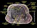
.svg.png) Anterior (frontal) view of the opened heart. White arrows indicate normal blood flow. | |
| Details | |
| Latin | atrium sinistrum |
| circumflex branch of left coronary artery | |
| oblique vein of the left atrium | |
| Identifiers | |
| Dorlands /Elsevier | a_71/12167887 |
| TA | A12.1.03.001 |
| FMA | 7097 |
| Anatomical terminology | |
The left atrium is one of four chambers in the heart. It receives oxygenated blood from the pulmonary veins, and pumps it into the left ventricle, via the mitral valve. Atria facilitate circulation primarily by allowing uninterrupted venous flow to the heart, preventing the inertia of interrupted venous flow that would otherwise occur at each ventricular systole.[1]
Structure


In the developing fetus there is an oval opening, the foramen ovale (heart) between the atria which normally closes at birth. If this does not close it could result in a hole in the heart, an atrial septal defect. In the fetus, the right atrium pumps blood into the left atrium, bypassing the pulmonary circulation (which is not needed in the fetus).
High in the upper part of the left atrium is an ear-shaped muscular pouch, the left atrial appendage (LAA). This is also called the auricle with reference to its shape. The atria themselves were previously called the auricles.
Function
The left atrium receives oxygenated blood from the pulmonary veins, and pumps it into the left ventricle, via the mitral valve (also known as the bicuspid valve). The atria facilitate circulation primarily by allowing an uninterrupted venous flow to the heart, preventing the inertia of interrupted venous flow that would otherwise occur at each ventricular systole.[1]
The left atrial appendage appears to "function as a decompression chamber during left ventricular systole and during other periods when left atrial pressure is high".[2]
Size
| Women | Men | |
|---|---|---|
| Diameter (mm) | 27–38 | 30–40 |
| Volume (ml) | 22–52 | 18–58 |
| Volume/BSA (ml/m²) | 16–28 | 16–28 |
| BSA, body surface area | ||
Blood supply
The left atrium is supplied mainly by the left circumflex coronary artery, though the branches are too small to be identified in a cadaveric human heart and are not named.[4]
The oblique vein of the left atrium is partly responsible for venous drainage; it derives from the embryonic left superior vena cava.
Clinical significance
In an adult, an atrial septal defect would result in the flow of blood in the reverse direction - from the left atrium to the right - which will reduce cardiac output, potentially causing cardiac failure and in severe or untreated cases cardiac arrest and sudden death.
In patients with atrial fibrillation, mitral valve disease, and other conditions, blood clots have a tendency to form in the LAA.[2] Blood clots caused by atrial fibrillation stem from the LAA in more than 90% of cases.[5] They may dislodge (forming emboli), which may lead to ischemic damage to the brain, kidneys, or other organs supplied by the systemic circulation.[6] Left atrial appendage occlusion is an experimental treatment to prevent stroke in atrial fibrillation.[7]
The left atrial appendage can serve as an approach for mitral valve surgery.[8] The left atrial appendage can be seen on a standard posteroanterior x-ray, where the lower level of the left hilum becomes concave.[9]
Other animals
Many other animals, including mammals, also have four-chambered hearts, which have a similar function. Some animals (amphibians and reptiles) have a three-chambered heart, in which the blood from each atrium is mixed in the single ventricle before being pumped to the aorta. In these animals, the left atrium still serves the purpose of collecting blood from the pulmonary veins.
References
- ↑ 1.0 1.1 Anderson, RM. The Gross Physiology of the Cardiovascular System (2nd ed., 2012) See "Chapter 1: Normal Physiology."
- ↑ 2.0 2.1 Al-Saady NM et al (1999) Left atrial appendage: structure, function, and role in thromboembolism: Review Heart 82:547-554
- ↑ Lang, RM; Bierig M, Devereux RB, Flachskampf FA, Foster E, Pellikka PA, Picard MH, Roman MJ, Seward J, Shanewise J, Solomon S, Spencer KT, St John Sutton M, Stewart W (March 2006). "Recommendations for chamber quantification". European Journal of Echocardiography 7 (2): 79–108. doi:10.1016/j.euje.2005.12.014. PMID 16458610. Retrieved 2012-08-26.
- ↑ Agur, Keith L. Moore, Arthur F. Dalley, Anne M.R. (2010). Clinically oriented anatomy (6th ed. ed.). Philadelphia: Wolters Kluwer Health/Lippincott Williams & Wilkins. p. 145. ISBN 9780781775250.
- ↑ Blackshear JL, Odell JA (February 1996). "Appendage obliteration to reduce stroke in cardiac surgical patients with atrial fibrillation". Ann. Thorac. Surg. 61 (2): 755–9. doi:10.1016/0003-4975(95)00887-X. PMID 8572814.
- ↑ Parekh A, Jaladi R, Sharma S, Van Decker WA, Ezekowitz MD (September 2006). "Images in cardiovascular medicine. The case of a disappearing left atrial appendage thrombus: direct visualization of left atrial thrombus migration, captured by echocardiography, in a patient with atrial fibrillation, resulting in a stroke". Circulation 114 (13): e513–4. doi:10.1161/CIRCULATIONAHA.106.617886. PMID 17000914.
- ↑ Stöllberger C, Schneider B, Finsterer J (December 2003). "Elimination of the left atrial appendage to prevent stroke or embolism? Anatomic, physiologic, and pathophysiologic considerations". Chest 124 (6): 2356–62. doi:10.1378/chest.124.6.2356. PMID 14665520.
- ↑ Guhathakurta S, Kurian VM, Manmohan, Cherian KM (2004). "Mitral valve reoperation through the left atrial appendage in a patient with mesocardia". Tex Heart Inst J 31 (3): 316–8. PMC 521780. PMID 15562857.
- ↑ Corne et al. (2002). Chest X-Ray Made Easy. Churchill Livingstone. ISBN 0-443-07008-3.
Additional images
-

Heart seen from above.
-

Base and diaphragmatic surface of heart.
-
Left atrium
See also
| ||||||||||||||||||||||||||||||||||||||||||||||||||||||||||||||||||||