Le Fort fracture of skull
| LeFort fracture | |
|---|---|
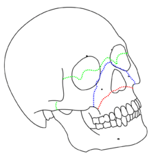 LeFort I (red), II (blue), and III (green) fractures | |
| Classification and external resources | |
| eMedicine | radio/385 |
Le Fort fractures are types of facial fractures involving the maxillary bone and surrounding structures in a usually bilateral and either horizontal, pyramidal or transverse way. LeFort fractures are classic in facial trauma. The Le Fort fracture was named after French surgeon René Le Fort (1869–1951.) Historically, Dr. Le Fort discovered these types of fracture patterns by examining break patterns of crush injuries of cadavers. [1]
Diagnosis
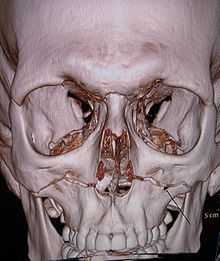
Diagnosis is made based physical exam findings with confirmation by axial CT. The patient is taken for radiography of the head and neck then after obvious fracture signs the patient is taken to CT scan for more specific anatomic information. To qualify for LeFort Fractures the pterygoid plates must be involved. These are seen posterior to the maxillary sinuses on axial CT and inferior to the orbital rim on coronal slices. Also, the palate is usually mobile on physical exam.
Classification


There are three types of Le Fort fractures:
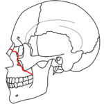
- Le Fort I fractures (horizontal) may result from a force of injury directed low on the maxillary alveolar rim in a downward direction. It is also known as a Guérin fracture or 'floating palate', and usually involves the inferior nasal aperture. The fracture extends from the nasal septum to the lateral pyriform rims, travels horizontally above the teeth apices, crosses below the zygomaticomaxillary junction, and traverses the pterygomaxillary junction to interrupt the pterygoid plates.
- Le Fort II fractures (pyramidal) may result from a blow to the lower or mid maxilla and usually involve the inferior orbital rim. Such a fracture has a pyramidal shape and extends from the nasal bridge at or below the nasofrontal suture through the frontal processes of the maxilla, inferolaterally through the lacrimal bones and inferior orbital floor and rim through or near the inferior orbital foramen, and inferiorly through the anterior wall of the maxillary sinus; it then travels under the zygoma, across the pterygomaxillary fissure, and through the pterygoid plates.


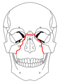
- Le Fort III fractures (transverse) are otherwise known as craniofacial dissociation and involve the zygomatic arch. These may follow impact to the nasal bridge or upper maxilla. These fractures start at the nasofrontal and frontomaxillary sutures and extend posteriorly along the medial wall of the orbit through the nasolacrimal groove and ethmoid bones. The thicker sphenoid bone posteriorly usually prevents continuation of the fracture into the optic canal. Instead, the fracture continues along the floor of the orbit along the inferior orbital fissure and continues superolaterally through the lateral orbital wall, through the zygomaticofrontal junction and the zygomatic arch. Intranasally, a branch of the fracture extends through the base of the perpendicular plate of the ethmoid, through the vomer, and through the interface of the pterygoid plates to the base of the sphenoid. This type of fracture predisposes the patient to CSF rhinorrhea more commonly than the other types.
Signs and Symptoms
Lefort I - Slight swelling of the upper lip, ecchymosis is present in the buccal sulcus beneath each zygomatic arch, malocclusion, mobility of teeth. Impacted type of fractures may be almost immobile and it is only by grasping the maxillary teeth and applying a little firm pressure that a characteristic grate can be felt which is diagnostic of the fracture. Percussion of upper teeth results in cracked pot sound. Guérin's sign is present characterised by ecchymosis in the region of greater palatine vessels.
Lefort II and Lefort III (common) - Gross edema of soft tissue over the middle third of the face, bilateral circumorbital ecchymosis, bilateral subconjunctival hemorrhage, epistaxis, CSF rhinorrhoea, dish face deformity, diplopia, enophthalmos, cracked pot sound.
Lefort II - Step deformity at infraorbital margin, mobile mid face, anesthesia or paresthesia of cheek.
Lefort III - Tenderness and separation at frontozygomatic suture, lengthening of face, depression of ocular levels, enophthalmos, hooding of eyes, tilting of occlusal plane with gagging on one side.
See also
References
- ↑ Allsop D, Kennett K (2002). "Skull and facial bone trauma". In Nahum AM, Melvin J. Accidental injury: Biomechanics and prevention. Berlin: Springer. pp. 254–258. ISBN 0-387-98820-3. Retrieved 2008-10-08.
External links
| Wikimedia Commons has media related to Fractures of the human maxilla. |
| ||||||||||||||||||||||||||||||||||||||||||||||||||||||||||||||||||||||||||||||||||||||||||||||||||