Lateralization of brain function
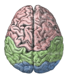
The longitudinal fissure separates the human brain into two distinct cerebral hemispheres, connected by the corpus callosum. The hemispheres exhibit strong, but not complete, bilateral symmetry in both structure and function. For example, structurally, the lateral sulcus generally is longer in the left hemisphere than in the right hemisphere, and functionally, Broca's area and Wernicke's area are located in the left cerebral hemisphere for about 95% of right-handers, but about 70% of left-handers.[1]
Broad generalizations are often made in "pop" psychology about one side or the other having characteristic labels, such as "logical" for the left side or "creative" for the right. These labels are not supported by studies on lateralization, as lateralization does not add specialized usage from either hemisphere.[2] Both hemispheres contribute to both kinds of processes,[3] and experimental evidence provides little support for correlating the structural differences between the sides with such broadly defined functional differences.[4]
The extent of any modularity, or specialization of brain function by area, remains under investigation. If a specific region of the brain, or even an entire hemisphere, is injured or destroyed, its functions can sometimes be assumed by a neighboring region in the same hemisphere or the corresponding region in the other hemisphere, depending upon the area damaged and the patient's age.[5] When injury interferes with pathways from one area to another, alternative (indirect) connections may develop to communicate information with detached areas, despite the inefficiencies.
Brain function lateralization is evident in the phenomena of right- or left-handedness[6] and of right or left ear preference,[7] but a person's preferred hand is not a clear indication of the location of brain function. Although 95% of right-handed people have left-hemisphere dominance for language, 18.8% of left-handed people have right-hemisphere dominance for language function. Additionally, 19.8% of the left-handed have bilateral language functions.[8] Even within various language functions (e.g., semantics, syntax, prosody), degree (and even hemisphere) of dominance may differ.[9]
Additionally, although some functions are lateralized, these are only a tendency. The trend across many individuals may also vary significantly as to how any specific function is implemented. The areas of exploration of this causal or effectual difference of a particular brain function include its gross anatomy, dendritic structure, and neurotransmitter distribution. The structural and chemical variance of a particular brain function, between the two hemispheres of one brain or between the same hemisphere of two different brains, is still being studied. Short of having undergone a hemispherectomy (removal of a cerebral hemisphere), no one is a "left-brain only" or "right-brain only" person.[10]
History of research on lateralization
Broca
One of the first indications of brain function lateralization resulted from the research of French physician Pierre Paul Broca, in 1861. His research involved the male patient nicknamed "Tan", who suffered a speech deficit (aphasia); "tan" was one of the few words he could articulate, hence his nickname. In Tan's autopsy, Broca determined he had a syphilitic lesion in the left cerebral hemisphere. This left frontal lobe brain area (Broca's area) is an important speech production region. The motor aspects of speech production deficits caused by damage to Broca’s area are known as Expressive aphasia. In clinical assessment of this aphasia, it is noted that the patient cannot clearly articulate the language being employed.
Wernicke
German physician Karl Wernicke continued in the vein of Broca's research by studying language deficits unlike expressive aphasia. Wernicke noted that not every deficit was in speech production; some were linguistic. He found that damage to the left posterior, superior temporal gyrus (Wernicke's area) caused language comprehension deficits rather than speech production deficits, a syndrome known as Receptive aphasia.
Advance in imaging technique
These seminal works on hemispheric specialization were done on patients and/or postmortem brains, raising questions about the potential impact of pathology on the research findings. New methods permit the in vivo comparison of the hemispheres in healthy subjects. Particularly, magnetic resonance imaging (MRI) and positron emission tomography (PET) are important because of their high spatial resolution and ability to image subcortical brain structures.
Movement and sensation
In the 1940s, neurosurgeon Wilder Penfield and his neurologist colleague Herbert Jasper developed a technique of brain mapping to help reduce side effects caused by surgery to treat epilepsy. They stimulated motor and somatosensory cortices of the brain with small electrical currents to activate discrete brain regions. They found that stimulation of one hemisphere's motor cortex produces muscle contraction on the opposite side of the body. Furthermore, the functional map of the motor and sensory cortices is fairly consistent from person to person; Penfield and Jasper's famous pictures of the motor and sensory homunculi were the result.
Split-brain patients
Research by Michael Gazzaniga and Roger Wolcott Sperry in the 1960s on split-brain patients led to an even greater understanding of functional laterality. Split-brain patients are patients who have undergone corpus callosotomy (usually as a treatment for severe epilepsy), a severing of a large part of the corpus callosum. The corpus callosum connects the two hemispheres of the brain and allows them to communicate. When these connections are cut, the two halves of the brain have a reduced capacity to communicate with each other. This led to many interesting behavioral phenomena that allowed Gazzaniga and Sperry to study the contributions of each hemisphere to various cognitive and perceptual processes. One of their main findings was that the right hemisphere was capable of rudimentary language processing, but often has no lexical or grammatical abilities.[11] Eran Zaidel, however, also studied such patients and found some evidence for the right hemisphere having at least some syntactic ability.
As stated above, language is primarily localized in the left hemisphere. One of the experiments carried out by Gazzaniga involved a split-brain patient sitting in front of a computer screen while having words and images presented on either side of the screen and the visual stimuli would go to either the right or left eye, and thus the left or right brain, respectively. It was observed that if a patient was presented with an image to his left eye (right brain), he would report not seeing anything. However, if he was able to feel around for certain objects, he could accurately pick out the correct object, despite not having the ability to verbalize what he saw. This led to confirmation that the left brain is localized for language while the right brain does not have this capability, and when the corpus callosum is cut and the two hemispheres cannot communicate for the speech to be produced.
For example: Patients with brain damage from surgery, stroke or infection sometimes develop a syndrome in which they can feel sensations in their hand, but they don't feel responsible for nor able to control its movements. In patients with a corpus callosotomy, alien hand syndrome most often manifests as uncontrolled but purposeful movements of the nondominant hand.
Sex differences
Sex differences are apparent in almost every aspect of neural anatomy and physiological psychology. This is also true with regards to lateralization differences between men and women. It is generally accepted that male brains are typically much more lateralized than female brains,[12][13] although this is challenged by a recent study.[2]
Lateralized cognitive processes
Language functions such as grammar, vocabulary and literal meaning[14][15] are typically lateralized to the left hemisphere, especially in right handed individuals.[15] While language production is left-lateralized in up to 90% of right-handed subjects, it is more bilateral, or even right lateralized in approximately 50% of left-handers.[16] In contrast, prosodic language functions, such as intonation and accentuation, often are lateralized to the right hemisphere of the brain.[17][18]
The processing of visual and auditory stimuli, spatial manipulation, facial perception, and artistic ability are represented bilaterally, but may show a right hemisphere superiority.[16] Numerical estimation, comparison and online calculation depend on bilateral parietal regions[19][20] while exact calculation and fact retrieval are associated with left parietal regions, perhaps due to their ties to linguistic processing.[19][20] Dyscalculia is a neurological syndrome associated with damage to the left temporo-parietal junction.[21] This syndrome is associated with poor numeric manipulation, poor mental arithmetic skill, and the inability to either understand or apply mathematical concepts.[22]
Depression is linked with a hyperactive right hemisphere, with evidence of selective involvement in "processing negative emotions, pessimistic thoughts and unconstructive thinking styles", as well as vigilance, arousal and self-reflection, and a relatively hypoactive left hemisphere, "specifically involved in processing pleasurable experiences" and "relatively more involved in decision-making processes".[23] Additionally, "left hemisphere lesions result in an omissive response bias or error pattern whereas right hemisphere lesions result in a commissive response bias or error pattern."[24] The delusional misidentification syndromes, reduplicative paramnesia and Capgras delusion are also often the result of right hemisphere lesions.[25][26] There is evidence[27] that the right hemisphere is more involved in processing novel situations, while the left hemisphere is most involved when routine or well rehearsed processing is called for.
Handedness and language
Broca's area and Wernicke's area are linked by a white matter fiber tract, the arcuate fasciculus. This axonal tract allows the neurons in the two areas to work together in creating vocal language. In more than 95% of right-handed men, and more than 90% of right-handed women, the left hemisphere is dominant in certain aspects of language and speech processing. In left-handed people, the incidence of left-hemisphere language dominance has been reported as 73%[28] and 61%,[8] suggesting left handed people tend to be less lateralized than right-handed people. In general, however, neuroimaging methods such as functional magnetic resonance imaging and magnetoencephalography show involvement of both hemispheres in many aspects of language processing, and the "dominance" of one hemisphere just refers to more brain activation relative to the other hemisphere (or better performance by that hemisphere on psycholinguistic tasks such as dichotic listening); it is not the case that language is "localized" in any one hemisphere laterally.
Methods of study
There are ways of determining whether particular cognitive functions tend to be lateralized to one cerebral hemisphere. The Wada Test introduces an anesthetic to one hemisphere of the brain via one of the two carotid arteries. Once the hemisphere is anesthetized, a neuropsychological examination is effected to determine whether cognitive functions such as language production, language comprehension, verbal memory, or visual memory are retained. Less invasive techniques, such as functional magnetic resonance imaging and transcranial magnetic stimulation may also be used to investigate the role of a particular cerebral hemisphere in a particular task, although these methods are costly. The divided visual field paradigm is another technique that has contributed to the study of hemispheric specialization.
Exaggeration of the lateralization concept
Terence Hines states that the research on brain lateralization is valid as a research program, though commercial promoters have applied it to promote subjects and products far outside the implications of the research.[29] For example, the implications of the research have no bearing on psychological interventions such as EMDR and neurolinguistic programming,[30] brain training equipment, or management training.[31]
Nonhuman brain lateralization
Specialization of the two hemispheres is general in vertebrates including fish, frogs, reptiles, birds and mammals with the left hemisphere being specialized to categorize information and control everyday, routine behavior, with the right hemisphere responsible for responses to novel events and behavior in emergencies including the expression of intense emotions. An example of a routine left hemisphere behavior is feeding behavior whereas as a right hemisphere is escape from predators and attacks from conspecifics.[32]
Advantages of brain lateralization
The widespread lateralization of many vertebrate animals indicates an evolutionary advantage associated with the specialization of each hemisphere.[33] In one experiment, baby chicks were lateralized before hatching by exposing their eggs to light.[34] These chicks were set to a task of picking out food from a bed of pebbles. Neither the lateralized, nor the non-lateralized chicks had a problem with this task, but the lateralized chicks only used the eye on the side of which they were lateralized to pick up the pebbles. When presented with a second task of watching for a cutout of a predatory hawk, the discrepancy between lateralized and non-lateralized chicks became evident. Lateralized chicks could pick food out of the pebbles with one eye and one half of the brain[35] while using the other eye and other half of their brain to monitor the skies for predators.[36] Not only could non-lateralized chicks not complete the two tasks simultaneously, but their performance of the single task deteriorated. This suggests that the evolutionary advantage of lateralization comes from the capacity to perform separate parallel tasks in each hemisphere of the brain.[33]
Additional images
-

Ventricles of brain and basal ganglia. Superior view. Horizontal section. Deep dissection
-
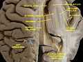
Ventricles of brain and basal ganglia. Superior view. Horizontal section. Deep dissection
See also
References
- ↑ Griggs, Richard A. Psychology: A Concise Introduction. p. 69.
- ↑ 2.0 2.1 Nielsen, Jared A., Brandon A. Zielinski, Michael A. Ferguson, Janet E. Lainhart, and Jeffrey S. Anderson. "An Evaluation of the Left-Brain vs. Right-Brain Hypothesis with Resting State Functional Connectivity Magnetic Resonance Imaging." PLOS ONE, 14 Aug. 2013. Web. 30 Aug. 2013.
- ↑ Westen et al. 2006 Psychology: Australian and New Zealand edition. John Wiley p.107
- ↑ Toga AW, Thompson PM (2003). "Mapping brain asymmetry". Nature Reviews Neuroscience 4 (1): 37–48. doi:10.1038/nrn1009. PMID 12511860.
- ↑ Pulsifer MB, Brandt J, Salorio CF, Vining EP, Carson BS, Freeman JM (2004). "The cognitive outcome of hemispherectomy in 71 children". Epilepsia 45 (3): 243–254. doi:10.1111/j.0013-9580.2004.15303.x. PMID 15009226.
- ↑ Knecht S, Dräger B, Deppe M, Bobe L, Lohmann H, Flöel A, Ringelstein EB, Henningsen H (2000). "Handedness and hemispheric language dominance in healthy humans". Brain : a journal of neurology 123 (12): 2512–2518. doi:10.1093/brain/123.12.2512. PMID 11099452.
- ↑ Schönwiesner M, Rübsamen R, von Cramon DY (2005). "Hemispheric asymmetry for spectral and temporal processing in the human antero-lateral auditory belt cortex". European Journal of Neuroscience 22 (6): 1521–1528. doi:10.1111/j.1460-9568.2005.04315.x. PMID 16190905.
- ↑ 8.0 8.1 Taylor, Insep and Taylor, M. Martin (1990) "Psycholinguistics: Learning and using Language". page 362
- ↑ Regarding different languages: http://www.bbc.co.uk/news/health-11181457
- ↑ Goswami U (2006). "Neuroscience and education: from research to practice?". Nat Rev Neurosci 7 (5): 406–11. doi:10.1038/nrn1907. PMID 16607400.
- ↑ Kandel E, Schwartz J, Jessel T. Principles of Neural Science. 4th ed. p1182. New York: McGraw–Hill; 2000. ISBN 0-8385-7701-6
- ↑ Maccoby, Eleanor (1974). The Psychology of Sex Differences. Stanford, California: Stanford University Press. ISBN 0804709742.
- ↑ Tomasi D, Volkow ND (June 2012). "Laterality Patterns of Brain Functional Connectivity: Gender Effects". Cerebral Cortex 22 (6): 1455–1462. doi:10.1093/cercor/bhr230. PMC 3450858. PMID 21878483.
- ↑ Boeree, C.G. (2004). "Speech and the Brain". Retrieved February 17, 2012.
- ↑ 15.0 15.1 Taylor, I. & Taylor, M. M. (1990). Psycholinguistics: Learning and using Language. Pearson. ISBN 978-0-13-733817-7. p. 367
- ↑ 16.0 16.1 Beaumont, J.G. (2008). Introduction to Neuropsychology, Second Edition. The Guilford Press. ISBN 978-1-59385-068-5. Chapter 7
- ↑ Ross ED, Monnot M (January 2008). "Neurology of affective prosody and its functional-anatomic organization in right hemisphere". Brain Lang. 104 (1): 51–74. doi:10.1016/j.bandl.2007.04.007. PMID 17537499.
- ↑ George MS, Parekh PI, Rosinsky N, Ketter TA, Kimbrell TA, Heilman KM, Herscovitch P, Post RM (July 1996). "Understanding Emotional Prosody Activates Right Hemisphere Regions". Arch Neurol. 53 (7): 665–670. doi:10.1001/archneur.1996.00550070103017. PMID 8929174.
- ↑ 19.0 19.1 Dehaene S, Spelke E, Pinel P, Stanescu R, Tsivkin S (May 1999). "Sources of mathematical thinking: behavioral and brain-imaging evidence" (PDF). Science 284 (5416): 970–4. doi:10.1126/science.284.5416.970. PMID 10320379.
- ↑ 20.0 20.1 Dehaene S, Piazza M, Pinel P, Cohen L (2003). "Three parietal circuits for number processing" (PDF). Cognitive Neuropsychology 20 (3–6): 487–506. doi:10.1080/02643290244000239. PMID 20957581.
- ↑ Levy LM, Reis IL, Grafman J (August 1999). "Metabolic abnormalities detected by 1H-MRS in dyscalculia and dysgraphia". Neurology 53 (3): 639–41. doi:10.1212/WNL.53.3.639. PMID 10449137.
- ↑ Dyscalculia Symptoms
- ↑ Hecht D (October 2010). "Depression and the hyperactive right-hemisphere". Neurosci. Res. 68 (2): 77–87. doi:10.1016/j.neures.2010.06.013. PMID 20603163.
- ↑ Braun CM, Delisle J, Guimond A, Daigneault R (March 2009). "Post unilateral lesion response biases modulate memory: crossed double dissociation of hemispheric specialisations". Laterality 14 (2): 122–64. doi:10.1080/13576500802328613. PMID 18991140.
- ↑ Devinsky O (January 2009). "Delusional misidentifications and duplications: right brain lesions, left brain delusions". Neurology 72 (1): 80–7. doi:10.1212/01.wnl.0000338625.47892.74. PMID 19122035.
- ↑ Madoz-Gúrpide A, Hillers-Rodríguez R (April 2010). "[Capgras delusion: a review of aetiological theories]". Rev Neurol 50 (7): 420–30. PMID 20387212.
- ↑ Goldberg, E. (2009). The New Executive Brain: Frontal Lobes in a Complex World. New York, NY: Oxford University Press. ISBN 978-0-19-532940-7.
- ↑ Knecht S, Dräger B, Deppe M, Bobe L, Lohmann H, Flöel A, Ringelstein EB, Henningsen H (2000). "Handedness and hemispheric language dominance in healthy humans". Brain 123 (12): 2512–2518. doi:10.1093/brain/123.12.2512. PMID 11099452.
- ↑ Hines, Terence (1987). "Left Brain/Right Brain Mythology and Implications for Management and Training". The Academy of Management Review 12 (4): 600–606. doi:10.2307/258066. JSTOR 258066.
- ↑ Drenth JD (2003). "Growing anti-intellectualism in Europe; a menace to science". Studia Psychologica 45 (1): 5–13., available in ALLEA Annual Report 2003, pp. 61–72
- ↑ Sala, Sergio Della (1999). Mind Myths: Exploring Popular Assumptions about the Mind and Brain. New York: Wiley. ISBN 0-471-98303-9.
- ↑ Vallortigara G, Rogers LJ (2005). "Survival with an asymmetrical brain: Advantages and disadvantages of cerebral lateralization". Behavioral and Brain Sciences 28 (4): 575–589; discussion 589–633. doi:10.1017/S0140525X05000105. PMID 16209828.
- ↑ 33.0 33.1 Halpern ME, Güntürkün O, Hopkins WD, Rogers LJ (2005). "Lateralization of the Vertebrate Brain: Taking the Side of Model Systems". The Journal of Neuroscience 25 (45): 10351–10357. doi:10.1523/JNEUROSCI.3439-05.2005. PMC 2654579. PMID 16280571.
- ↑ Rogers LJ (1990). "Light Input and the Reversal of Functional Lateralization in the Chicken Brain". Behav Brain Res 38 (3): 211–21. doi:10.1016/0166-4328(90)90176-F. PMID 2363841.
- ↑ Deng C, Rogers LJ (1997). "Differential Contributions of the Two Visual Pathways to Functional Lateralization in Chicks". Behav Brain Res 87 (2): 173–82. doi:10.1016/S0166-4328(97)02276-6. PMID 9331485.
- ↑ Rogers LJ (2000). "Evolution of Hemispheric Specialization: Advantages and Disadvantages". Brain Lang 73 (2): 236–53. doi:10.1006/brln.2000.2305. PMID 10856176.
Further reading
- Harnad, Stevan; Doty, R.W., Goldstein, L., Jaynes, J. & Krauthamer, G. (1977). Lateralization in the nervous system. Academic Press. ISBN 978-0-12-325750-5.
- Luria, A. R. (1966). Higher cortical functions in man. Basic Books.
- Ornstein, Robert (1998). The Right Mind: Making Sense of the Hemispheres. Harcourt Brace International. ISBN 978-0-15-600627-9.
- Drenth, Pieter (2006). Walks in the Garden of Science: Selected Papers and Lectures (PDF). Conference allea.
- Josse G, Tzourio-Mazoyer N (2003). "Review: Hemispheric specialization for language". Brain Research Reviews 44 (1): 1–12. doi:10.1016/j.brainresrev.2003.10.001. PMID 14739000.
| Laterality | |||
|---|---|---|---|
| Side | Left | Both | Right |
| General | Ambidexterity | ||
| In cognitive abilities | Geschwind–Galaburda hypothesis | ||
| In brain | |||
| In eyes | Ocular dominance | ||
| In hands | Left-handedness | Cross-dominance | Right-handedness |
| Handedness in boxing | Southpaw stance | Orthodox stance | |
| Handedness in people | |||
| Handedness related to | |||
| Handedness measurement | Edinburgh Handedness Inventory | ||
| Handedness genetics | LRRTM1 | ||
| In heart | Levocardia | Dextrocardia | |
| In major viscera | Situs solitus | Situs ambiguus | Situs inversus |
| In feet | Footedness | ||